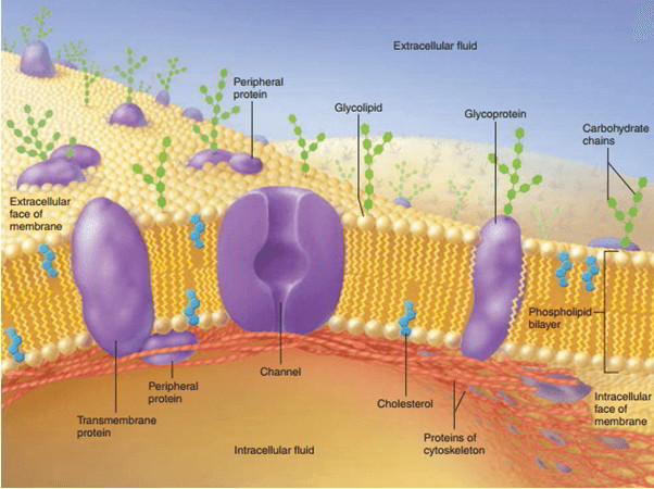
The cell membrane is a phospholipid bi-layer into which proteins, glycoproteins, and glycolipids are ingrained. The cell membrane is also known as plasma membrane or plasmalemma. This membrane separates the fluid outside the cell called extracellular fluid (ECF) and the fluid inside the cell called intracellular fluid (ICF). The cell membrane is a semipermeable membrane. So, there is complimentary exchange of particular substances in between ECF and ICF. Density of the cell membrane differs from 75 to 111 Å. The phospholipid bi-layer supplies a barrier to diffusion. The lipid slows down movement of ions and other charged molecules.
On the other hand, lipid-soluble substances quickly scattered throughout the membrane however, have trouble taking a trip through the aqueous extracellular and intracellular fluid Lipophilic molecules can collect in the interior of cell membranes Little polar molecules, such as water and urea, quickly scattered throughout the membrane, facilitated by selective channels that cover the lipid bilayer. Diffusion of glucose and other big polar molecules is hampered by the plasma membrane, and cellular uptake of glucose needs particular transport proteins.
Proteins in the cell membrane selectively manage the cellular entry and exit of water-soluble, however not lipid-soluble, products. Movement throughout the membrane can happen passively down a concentration gradient (from high to low concentration) or by active transport versus the concentration gradient (from low to high). Active transport processes need energy.
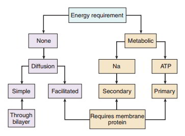
Diffusion takes place down a concentration gradient. The efficiency of diffusion is increased by increasing the concentration gradient, increasing the permeability, increasing
the surface area, or reducing the range over which the substance should take a trip
Cell membranes display a diplomatic immunity of facilitated diffusion. This transport process permits the transmembrane movement of substances that are badly soluble in the phospholipid bilayer. Glucose absorption throughout the intestinal epithelia highlights both secondary active transport and facilitated diffusion. On the apical surface, glucose goes into the cell by a secondary active transport process, combined to Na+ entry. This process permits glucose uptake even when the extracellular luminal glucose concentration is lower than the intracellular glucose concentration. Glucose exits the cell on the basolateral surface by facilitated diffusion. No energy is used up, and the glucose moves down the concentration gradient. The net result is that glucose is soaked up from the lumen of the intestine into the body. Movement throughout the basolateral surface takes place by transport proteins, however, no energy (besides the glucose concentration gradient) is included. The dependence on transport proteins, nevertheless, indicates that substances moving by facilitated diffusion program saturation kinetics, where the variety of transport proteins can restrict the optimum rate of the substance.
In a totally random world, diffusion would make sure the even circulation of all substances Life, nevertheless, depends upon the development, maintenance, and usage of concentration gradients. The development of concentration gradients can not happen by diffusion. The energy needed to move solutes versus their concentration gradient originates from hydrolysis of ATP in main active transport or from energy originated from a pre-existing concentration gradient in secondary active transport.
Composition of Cell Membrane
Cell membrane is made up of 3 types of substances:
- Proteins (55%).
- Lipids (40%).
- Carbohydrates (5%).
Structure of Cell Membrane
On the basis of structure, cell membrane is called a unit membrane or a three-layered membrane. The electron microscopic study exposes 3 layers of cell membrane, specifically, one central electron-lucent layer and 2 electron-dense layers. The 2 electron-dense layers are positioned one on either side of the central layer. The central layer is a lipid layer formed by lipid substances. The other 2 layers are protein layers formed by proteins cell membrane which consists of some carbohydrate molecules too.
Structural Model of the Cell Membrane
- Danielli-Davson Model: It was the very first proposed standard model of membrane structure It was proposed by James F Danielli and Hugh Davson in 1935 and it was accepted by researchers for several years. This model was generally a ‘sandwich of lipids’ covered by proteins on both sides.
- Unit Membrane Model: In 1957, JD Robertson changed ‘DanielliDavson model’ to ‘Unit membrane model’ on the basis of electron microscopic research studies.
- Fluid Mosaic Model: Later on in 1972, SJ Vocalist and GL Nicholson proposed ‘The fluid mosaic model’. According to them, the membrane is a fluid with mosaic of proteins (mosaic ways pattern formed by plan of various colored pieces of stone, tile, glass or other such products). This model is accepted by the researchers till now. In this model, the proteins are discovered to drift in the lipid layer rather of forming the layers of the sandwich-type model.
Lipid Layers of the Cell Membrane
The central lipid layer is a bi-layered structure which is formed by a thin film of lipids. The particular function of lipid layer is that, it is fluid in nature and not a strong structure So, the parts of the membrane move from one point to another point along the surface of the cell. The products liquified in lipid layer likewise move to all locations of the cell membrane.
Significant lipids are:
- Phospholipids
- Cholesterol
Phospholipids
Phospholipids are the lipid substances consisting of phosphorus and fats. Aminophospholipids, sphingo – myelins, phosphatidylcholine, phosphatidylethanolamine, phosphatidylglycerol, phosphatidylserine and phospha tidylinositol are the phospholipids present in lipid layer of cell membrane. Phospholipid molecules are set up in 2 layers. Each phospholipid molecule looks like the headed pin fit. The external part of the phospholipid molecule is called the head part and the inside is called the tail part. Head part is the polar end and it is soluble in water and has strong affinity for water (hydrophilic). Tail part is the non-polar end. It is insoluble in water and fended off by water (hydrophobic). 2 layers of phospholipids are set up in such a method that the hydrophobic tail parts satisfy in the centre of the membrane. Hydrophilic head parts of external layer face the ECF and those of the inner layer face ICF (cytoplasm).
Cholesterol
Cholesterol molecules are set up between the phospholipid molecules. Phospholipids are soft and oily structures and cholesterol assists to load the phospholipids in the membrane. So, cholesterol is accountable for the structural stability of lipid layer of the cell membrane.
Functions of Lipid Layer in Cell Membrane
Lipid layer of the cell membrane is a semipermeable membrane and permits just the fat-soluble substances to go through it. Hence, the fat-soluble substances like oxygen, co2 and alcohol can go through this lipid layer. The water-soluble substances such as glucose, urea and electrolytes can not go through this layer.
Protein Layers of the Cell Membrane
Protein layers of the cell membrane are electron-dense layers. These layers cover 2 surfaces of the central lipid layer. Protein layers offer security to the central lipid layer. The protein substances present in these layers are primarily glycoproteins. Protein molecules are categorized into 2 classifications:
1.Important proteins or transmembrane proteins are the proteins that go through whole density of cell membrane from one side to the opposite. These proteins are securely bound with the cell membrane.
Examples of essential protein: i. Cell adhesion proteins ii. Cell junction proteins iii. Some provider (transport) proteins iv. Channel proteins v. Some hormone receptors vi. Antigens vii. Some enzymes.
2.Peripheral proteins or peripheral membrane proteins are the proteins which are partly embedded in the external and inner surfaces of the cell membrane and do not permeate the cell membrane. Peripheral proteins are loosely bound with essential proteins or lipid layer of cell membrane. So, these protein molecules dissociate easily from the cell membrane. Examples of peripheral proteins: i. Proteins of cytoskeleton ii. Some provider (transport) proteins iii. Some enzymes.
Functions of Proteins in Cell Membrane
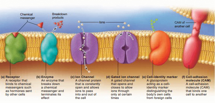
- Receptors. Much of the chemical signals by which cells interact (epinephrine, for instance) can not get in the target cell however, bind to surface proteins called receptors. Receptors are normally particular for one specific messenger, similar to an enzyme that specifies for one substrate.
- Second-messenger systems When a messenger binds to a surface receptor, it might activate modifications within the cell that produce a 2nd messenger in the cytoplasm. This process includes both transmembrane proteins (the receptors) and peripheral proteins.
- Enzymes. Enzymes in the plasma membrane perform the lasts of starch and protein digestion in the little intestine, assistance produce 2nd messengers, and break down hormones and other signalling molecules whose task is done, hence stopping them from exceedingly promoting a cell.
- Ion channels Ion channels are transmembrane proteins or protein clusters with pores that permit water and liquefied ions to go through the membrane. Some channels are constantly open, while others are gates that open and close under various situations, hence identifying when solutes can pass. These gates react to 3 types of stimuli: ligand-regulated gates react to chemical messengers, voltage-regulated gates to modifications in electrical potential (voltage) throughout the plasma membrane, and mechanically managed gates to physical tension on a cell, such as stretch and pressure.
- By managing the movement of electrolytes through the plasma membrane, gated channels play a crucial role in the timing of nerve signals and muscle contraction.
- Providers. Providers are trans-membrane proteins that bind to glucose, electrolytes, and other solutes and move them to the opposite of the membrane. Some providers, called pumps, take in ATP in the process.
- Cell-identity markers. Glycoproteins add to the glycocalyx, a carbohydrate surface finish. To name a few functions, this imitates a “recognition tag” that allows our bodies to inform which cells come from it and which are foreign intruders.
- Cell-adhesion molecules. Cells abide by one another and to extra cellular product through membrane proteins called cell-adhesion molecules (Webcams). With couple of exceptions (such as blood cells and metastasizing cancer cells), cells do not grow or make it through typically unless they are mechanically connected to the extracellular product. Unique occasions such as sperm-egg binding and the binding of an immune cell to a cancer cell likewise need Webcams.
Second Messengers
Second messengers are of such significance that they need a closer appearance. You will discover this info important for your later understanding of hormone and neurotransmitter action. Let’s try and understand how the hormone epinephrine promotes a cell. Epinephrine, the “very first messenger” can not go through the plasma membrane, so it binds to a surface receptor. The receptor is connected on the intracellular side to a peripheral protein called a G-protein. G proteins are called for the ATP-like chemical, guanosine triphosphate (GTP), from which they get their energy.
When triggered by the receptor, a G protein passes on the signal to another membrane protein, adenylate. Adenylate cyclase eliminates 2 phosphate groups from ATP and transforms it to cyclic AMP (cAMP), the 2nd messenger. Cyclic AMP then triggers enzymes called kinases in the cytosol. Kinases include phosphate groups to other cellular enzymes. This triggers some enzymes and shuts down others, however, in either case, it sets off a fantastic range of physiological modifications within the cell. As much as 60% of presently utilized drugs work by modifying the activity of G proteins.
Carbohydrates of the Cell Membrane
A few of the carbohydrate molecules present in cell membrane are connected to proteins and form glycoproteins (proteoglycans). Some carbohydrate molecules are connected to lipids and form glycolipids. Carbohydrate molecules form a thin and loose covering over the whole surface of the cell membrane called glycocalyx.
Functions of Carbohydrates in Cell Membrane
- Carbohydrate molecules are adversely charged and do not allow the adversely charged substances to move in and out of the cell.
- Glycocalyx from the neighbouring cells assists in the tight fixation of cells with one another.
- Some carbohydrate molecules function as the receptors for some hormones.
Functions of Cell Membrane
- Protective function: Cell membrane safeguards the cytoplasm and the organelles present in the cytoplasm
- Selective permeability: Cell membrane serves as a semipermeable membrane, which permits just some substances to go through it and serves as a barrier for other substances
- Absorptive function: Nutrients are soaked up into the cell through the cell membrane
- Excretory function: Metabolites and other waste products from the cell are excreted out through the cell membrane
- Exchange of gases: Oxygen goes into the cell from the blood and co2 leaves the cell and goes into the blood through the cell membrane
- Maintenance of sizes and shape of the cell: Cell membrane is accountable for the maintenance of sizes and shape of the cell.

 (58 votes, average: 4.55 out of 5)