The process of bone formation is called ossification (os-i-fi-ka’-shun). It begins during the sixth or seventh week of embryonic development. Bones are formed by the replacement of existing connective tissues with bone. There are two types of bone formation: intramembranous ossification and endochondral ossification.
In both types of ossification, some primitive connective tissue cells are changed into bone-forming cells called osteoblasts (os’-te-o-blasts). Osteoblasts deposit bone matrix around themselves and soon become imprisoned in lacunae. Once this occurs, they are called osteocytes.
Intramembranous Ossification
Most skull bones are formed by intramembranous ossification. Connective tissue membranes form early in embryonic development at sites of future intramembranous bones. Later, some connective tissue cells become osteoblasts and deposit spongy bone within the membranes starting in the centre of the future bone. Osteoblasts from the periosteum deposit a layer of compact bone over the spongy bone.
Some bone must be removed and re-formed in order to produce the correct shape of the bone as it develops and grows. Cells that remove bone matrix are called osteoclasts. The opposing actions of osteoblasts and osteoclasts ultimately produce the shape of the mature bone.
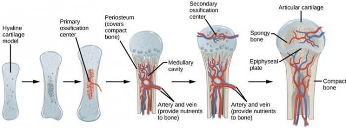
Endochondral Ossification
Most bones of the body are formed by endochondral (en-do-kon’-drul) ossification. Future endochondral bones are preformed in hyaline cartilage early in embryonic development. The figure above illustrates the ossification of a long bone.
In long bones, a new periosteum develops around the diaphysis of the hyaline cartilage template. Osteoblasts from the periosteum form a collar of compact bone around the diaphysis. A primary ossification center also forms in the middle of the cartilage shaft due to the enlargement of chondrocytes and a loss of cartilage matrix between lacunae. Calcification, which involves the depositing of calcium salts, occurs within the primary ossification center and leads to the death of chondrocytes. Blood vessels and nerves penetrate into the primary ossification center carrying along osteoblasts from the periosteum. As secondary ossification centers form in the epiphyses of the cartilage template, osteoclasts begin to remove spongy bone from the diaphysis to form the medullary cavity. The bone continues to grow as ossification progresses. As cartilage continues to be replaced, the cartilage between the primary and secondary ossification centers decreases until only a thin plate of cartilage, the epiphysial plate, separates the epiphyses from the diaphysis.
Subsequent growth in diameter results from the continued formation of compact bone by osteoblasts from the periosteum. Growth in length occurs as bone replaces cartilage on the diaphysis side of each epiphysial plate while new cartilage is formed on the epiphysis side. The opposing actions of osteoblasts and osteoclasts continually reshape the bone as it grows.
Growth usually continues until about age 25, when the epiphysial plates are completely replaced by bone. After this, growth in the length of a bone is not possible. The visible lines of fusion between the epiphyses and the diaphysis are called epiphysial lines.
Comparison of Intramembranous And Endochondral Ossification
| Intramembranous | Endochondral | ||
| 1. | Membranes of embryonic connective tissue form at sites of future bones. | 1. | Bone is preformed in hyaline cartilage. |
| 2. | Some connective tissue cells become osteoblasts, which deposit spongy bone within the membrane. | 2. | Osteoblasts of periosteum form a collar of compact bone that thickens and grows toward each end of the bone. |
| 3. | Osteoblasts from the enclosing membrane, now called the periosteum, deposit a layer of compact bone over the spongy bone. | 3. | Cartilage is calcified, and osteoblasts derived from the periosteum form spongy bone, which replaces cartilage in ossification centers. The spongy bone is later removed in the diaphysis to form the medullary cavity. |
Homoeostasis of Bone
Bones are dynamic, living organs, and they are continually restructured throughout life. This occurs by the removal of bone matrix by osteoclasts and by the deposition of new bone matrix by osteoblasts. Physical activity causes the density and volume of bones to be maintained or increased, though inactivity results in a reduction in bone density and volume.
Calcium salts may be removed from bones to meet body needs anytime blood calcium levels are low, such as when dietary intake is inadequate. When dietary calcium intake increases blood calcium to a sufficient level, some calcium is used to form new bone matrix.
Children have a relatively large number of protein fibers in their bone matrix, which makes their bones somewhat flexible. But as people age, the amount of protein gradually decreases. This trend causes older people to have brittle bones that are prone to fractures. Older persons may also experience a gradual loss of bone matrix, which reduces the strength of the bones. A severe reduction in bone density, and therefore increased risk of fracture, is called osteoporosis.

 (48 votes, average: 4.63 out of 5)