The technique of magnetic resonance imaging was used since the early 1980s to supply exceptionally detailed sectional pictures of internal organs and structures. These pictures are created by a computer using advice received from a scanner. Unlike X rays or computerized tomography (CT) scanning, MRI doesn’t involve you being exposed to potentially dangerous radiation: rather, it uses magnets and radio waves.
Pictures from MRI are much like those generated by CT scan, but MRI can differentiate unusual tissue considerably more definitely. MRI scans may also be shot at a greater range of planes through the body than is possible with CT scan and thus can be used to picture any part of the body. MRI is radiation-free and is
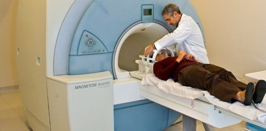
Regarded as among the safest imaging techniques accessible. During an MRI scan, you lie inside a scanner encompassed by a big, strong magnet.
A receiving magnet is subsequently put around the part of your body which is to be inquired. If big regions, including the abdomen, are to be imaged, the receiving magnet is fitted inside the MRI scanner; for smaller regions, including a joint, a magnet may be set around the part to be scanned. The MRI scanner is used from an adjoining room because the computer controlling the scanner must be shielded from the strong magnetic field created during the process. MRI scanners can be quite noisy and you might be given earplugs or headphones to wear. Each MRI scan continues just a couple of minutes, but a complete assessment may need many scan sequences and may last between a quarter hour and an hour. If you’re feeling nervous about the process you might be given a light sedative; you can also be asked if you’d like to bring a relative or a friend for moral support.
MRI scan is particularly useful for looking at the brain and for finding brain tumours. MRI is, in addition, valuable for looking at the spinal cord and may be used to inquire low back pain. Sports harms, particularly in the knee, are increasingly being analyzed using MRI. In a few instances, MRI was used to examine the breasts. MRI scans reveal the places of tumours within the breast tissue more precisely than simple two dimensional X rays. Furthermore, since MRI will not use radiation, MRI scans can be replicated frequently, enabling physicians to monitor a state attentively.

 (55 votes, average: 4.65 out of 5)