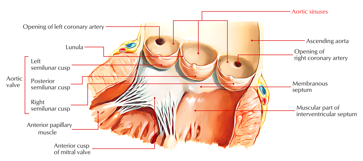
The aortic valve sinuses are located just before expansion of the wall of the ascending aorta, one over each of the distinct leaflets of the valve. The luminal diameter is enlarged and the thickness of the wall is reduced in relation to the regions that are located directly superiorly as well as inferiorly inside the area of the sinuses.

Aortic Sinuses
Features
- Functionally, the sinuses are important in maintaining the normal flow of blood into the coronary ostia and also the unblocked state of the valve leaflets during systole.
- A thickening called the supravalvar ridge marks the superior margin of each sinus.
- In the fully open aortic valve, the ridge is located superior towards the upper margin of the leaflets.
- The ostia of the coronary arteries are located just inferior towards the ridge of the left and right leaflets.
- The quantity of fibrous tissue inside the sinus is reduced as the quantity of elastic tissue is raised while rising superiorly.
- Compared to those of the pulmonary trunk, the aortic sinuses are more prominent.
Structure
- The interface in the middle of the LV and the ascending aorta, spreading out from the sinotubular junction in the aorta towards the basal valvular leaflets with their connection within the LV is referred to as the aortic root.
- Approximately two-thirds of the circumference of the lower part of the aortic root is connected to the muscular ventricular septum, with the remaining one-third in fibrous continuity with the aortic leaflet of the mitral valve.
- Its constituents are the sinuses of Valsalva, the fibrous interleaflet triangles, along with the valvular leaflets themselves.
- Three symmetric, semilunar-shaped cusps create the aortic valve. The indentation of each cusp is called the “sinus of Valsalva.”
- The aortic cusps are strongly attached inside the root of the aorta to the fibrous skeleton.
- A circular rim on the innermost side of the aortic wall, at the upper margin of each sinus, is the sinotubular rim that is the junction of the sinuses and the aorta.
- According to their location in the body, the aortic cusps are named left cusp and right cusp, both of them anteriorly facing the pulmonic valve and posterior cusp.
- The left coronary artery is produced by the left aortic sinus and the right coronary artery is produced by the right aortic sinus.
- Generally there are no vessels that originate from the posterior aortic sinus, so it is known as the non-coronary sinus.
- The intersections of the sinuses of Valsalva are marked by three equally spaced locations of minimal binding within the aortic root.
- The junctions in the middle of the adjacent sinuses are associated with the intersections among the aortic valve leaflets, while each sinus is related to a leaflet of the aortic valve.
Clinical Significance
- Sinus of Valsalva aneurysm (SOVA), a hereditary or acquired cardiac defect that is very rare and presents itself often during cardiac imaging as a secondary discovery.
- Even though an echocardiogram is the regular imaging technique for such discoveries, cardiac computed tomography angiography (CCTA) is more progressively used.
- If SOVA is diagnosed, CCTA is also a useful test for patients before surgical repair who are at low to intermediate level risk for coronary artery disease (CAD).
- The need for invasive angiography is removed in most cases because CCTA can precisely point out CAD, which may be more dangerous in SOVA patients since their coronaries may be more difficult to involve and their aortic root may be more susceptible to injury.
- Even though the treatment of choice previously has been surgery, the transcatheter methods have contributed to the range of nonsurgical alternatives for healing.


