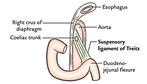
The attachment of the suspensory muscle of the duodenum (ligament of Treitz) supports the Duodenojejunal junction. The angle of the duodenojejunal flexure expands by contraction of this suspensory muscle, enhancing motion of the intestinal tract contents. A thin layer of skeletal muscle via the diaphragm and a fibromuscular band of smooth muscle via the 3rd and 4th parts of the duodenum are the typical constituents of the suspensory muscle of the duodenum.
Origin
- The superior muscular part emerges as a layer of muscle from the right esophageal crus of the respiratory diaphragm, along with muscular and ligamentous fibers emerging around origin of the celiac trunk and superior mesenteric artery and is skeletal (voluntary) muscle.
- It is created by a tendon having two muscular ends of different embryological genesis and function, because way the muscle has an uncommon structure.
- The inferior section of the muscle has actually been described as constant with both of the longitudinal and rounded muscle layer of the intestine at the duodenojejunal junction and is smooth (involuntary) muscle.

Ligament of Treitz
Insertion
- Posterior to the pancreas and splenic vein and anterior to the left renal vein, is the area where the suspensory muscle goes through.
- The origin of the intraperitoneal jejunum is marked by the Ligament of Treitz or the suspensory muscle of the duodenum
- This ligament exists about 75% of the time.
- Smooth muscle at the duodenojejunal flexure mixes with a strap of smooth muscle ranging from the connective tissue over the celiac artery and right diaphragmatic crus.
- It serves as a ligament to support the duodenojejunal flexure, however has very little importance as a muscle.
Structure
- A fold of peritoneum over the suspensory muscle of the duodenum creates the ligament of Treitz. This muscle is likewise called the “muscle of Treitz” or “musculus suspensorius duodenii”.
- The ligament of Treitz is consisted of two parts,.
- The first which passes from the right crus of the diaphragm to connective tissue surrounding the coeliac artery.
- The second, muscular, part comes down from stated connective tissue to the duodenum, in between the pancreas and the left renal vein. It is the second part that in fact suspends the duodenojejunal flexure.
- It is in fact a non-striated muscle which by the connective tissue around the coeliac artery and left crus of the diaphragm, is connected to the diaphragm near the oesophageal opening, which comes down to be placed in the distal duodenum and the duodeno-jejunal flexure (40%). It is made up of a fibromuscular band of smooth muscle via the third and fourth parts of the duodenum. A surrounding slip of skeletal muscle via the diaphragm is typically connected with benefit however unique anatomically.
Function
- Contraction of this muscle expands the angle of the duodenojejunal flexure, enhancing motion of the intestinal contents.
- The suspensory muscle enter posterior to the pancreas and splenic vein and anterior to the left renal vein.
Clinical Significance
The ligament of Treitz is site:
- to get the radiological diagnosis of intestinal tract malrotation and incomplete rotation.
- for judging between lower and upper gastrointestinal bleeding.
Short ligament of Treitz causes congenital superior masenteric artery syndrome.

 (56 votes, average: 4.48 out of 5)