All the living things are made up ofcells The human body is made up of about 75 trillion cells, the tiniest living systems that exist. Body cells can be categorized into about 300 types, such as neurons, epithelial cells, muscle cells, and red bloodcells Each type of cell has a special structure for carrying out particular functions Although these cells differ in size, shape, and function, they display numerous structural and practical resemblances.
Human cells are extremely little and show up just with a microscope Understanding of cell structure is based mostly on the assessment of cells with an electron microscope, a type of microscope that supplies zooms as much as 200,000× or more.
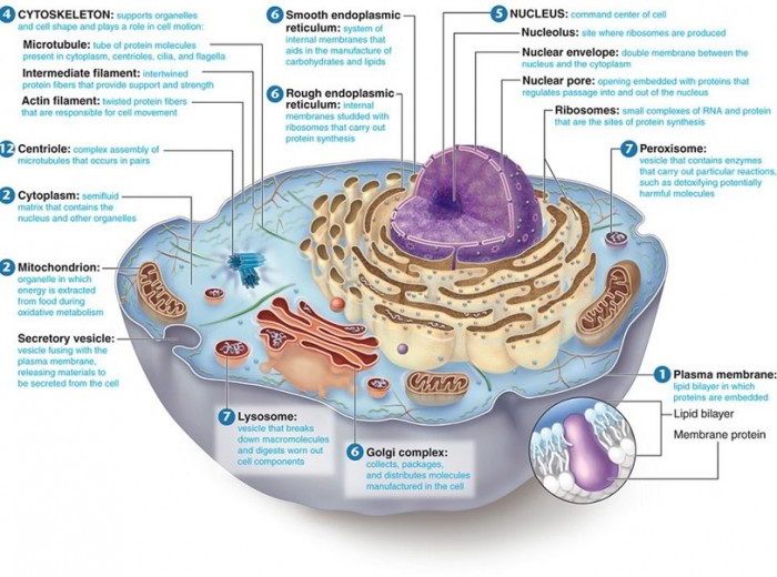
Although human cells are little, they are incredibly complex with numerous specialized parts The composite cell in figure highlights the significant structures understood to make up human cells These structures are revealed as they appear in electron microscope images. A lot of, however not all, of these structures are discovered in each human cell. The 3 typical parts discovered in all the cells are the plasma membrane, cytoplasm, and nucleus The other structures may or may not exist, depending upon cell type. As each part of a cell is gone over, note its structure and relationship to other structures in figure listed below.
Characteristics of Cell Each cell in the body
1. Needs nutrition and oxygen
2. Produces its own energy essential for its growth, repair work and other activities
3. Removes co2 and other metabolic wastes
4. Keeps the medium, i.e. the environment for its survival Cell
5. Shows instant response to the entry of intruders like bacteria or poisonous substances into the body
6. Reproduces by division. There are some exceptions like neuron, which do not reproduce.
Structure of a Cell
The Plasma Membrane
The plasma membrane forms the external boundary of a cell. It keeps the stability of the cell and separates the intracellular fluid from the extracellular fluid surrounding the cell. The plasma membrane includes 2 layers of phospholipid molecules, lined up back-to-back, with their fat tails forming the internal layer of the membrane and their polar heads dealing with the extracellular and intracellular fluids.
Cholesterol molecules are spread amongst the phospholipids, where they serve to increase the stability of the plasma membrane. The fat tails of the plasma membrane permit lipid-soluble substances to pass throughout the membrane however, avoid the passage of water-soluble substances Therefore, the plasma membrane works as a barrier in between water-soluble substances in the intracellular and extracellular fluids.
Various types of protein molecules are embedded in the plasma membrane, and each type has particular functions Some proteins form channels or pores through which water and water-soluble substances move throughout the membrane. A few of these proteins permit a variety of substances to pass across; others allow just particular molecules or ions to go into or leave a cell. Some proteins work as receptors for substances, such as hormones, that affect the function of a cell. Other proteins are enzymes that catalyse metabolic responses.
Specific proteins, in mix with carbohydrate molecules, work as recognition markers enabling cells to acknowledge each other. These recognition markers permit the lymphoid system to acknowledge “self” cells from “non-self” (foreign) cells, a difference necessary in combating pathogens All products that go into or leave a cell needs to pass throughout the plasma membrane. The plasma membrane is a selectively permeable membrane since it enables just specific substances to go into or leave the cell. Whether a substance can pass throughout the membrane is identified by a variety of aspects that consist of the substance’s size, solubility, electrical charges, and accessory to provider proteins.
Cytoplasm
The interior of a cell in between the plasma membrane and the nucleus is filled with a semifluid product called cytoplasm. It is made up of a gel-like fluid called cytosol, which is 75–90% of water and includes natural and inorganic substances, and little subcellular structures referred to as organelles.
Organelles
A variety of organelles, or little organs, are surrounded by cytosol. Organelles are identified by size, shape, structure, and particular function.
| Organelles | Functions |
| Rough endoplasmic reticulum |
|
| Smooth endoplasmic reticulum |
|
| Golgi apparatus | 1. Processing, product packaging, labelling and shipment of proteins and lipids |
| Lysosomes |
|
| Peroxisomes |
|
| Centrosome | 1. Movement of chromosomes throughout cell division |
| Mitochondria |
|
| Ribosomes | 1. Synthesis of proteins |
| Cytoskeleton |
|
| Nucleus |
|
Nucleus
The biggest organelle is the nucleus, a round or egg-shaped structure that is somewhat denser than the surrounding cytoplasm. It is separated from the cytoplasm by a double-layered nuclear envelope including many pores that permit the movement of products in between the nucleus and cytoplasm.
- Nuclear membrane
The nuclear membrane is double layered permeable structure having a 40,270nm large space called perinuclear tank which is constant with the lumen of endoplasmic reticulum. The external layer of the nuclear membrane is constant with endoplasmic reticulum. The exchange of products in between the nucleoplasm and cytoplasm happens through the nuclear membrane.
- Nucleoplasm The nucleoplasm or the nuclear matrix is a gel-like ground substance including a big amount of genetic product in the form of DNA. When a cell is not dividing, the nucleoplasm looks like dark staining thread-like product called nuclear chromatin. Throughout cell division, the chromatin product is transformed into rod-shaped structures, the chromosomes. There are 46 chromosomes (23sets) in all the dividing cells of the body other than the gamete (sex cells) which include just 23 chromosomes (haploid number). Each chromosome is made up of 2 chromatids linked at the centromere to form ‘X’ setup having variation of the area of centromere. number). Each chromosome is made up of 2 chromatids linked at the centromere to form ‘X’ setup having variation of the area of centromere. The chromosomes are made up of 3 components: DNA, RNA and other nuclear proteins The nuclear DNA brings the genetic info which is passed through RNA into the cytoplasm for synthesis of proteins of comparable composition.
- Nucleolus The nucleus might include several rounded bodies called nucleoli. The nucleoli are the site of synthesis of ribosomal RNA. The nucleoli are more typical in growing cells or in cells that actively synthesise proteins.
Chromosomes
The most essential structures within the nucleus, include DNA and proteinsThe DNA of chromosomes includes coded directions, called genes, that identify the functions of the cell. When a cell is not dividing, chromosomes are reached form thin threads that look like chromatin granules when seen microscopically. Throughout cell division, the chromosomes coil, reduce, and end up being rod-shaped Each human body cell includes 23 sets of chromosomes, with an overall of 46 in all. Several thick round bodies, called the nucleolus or nucleoli are likewise present in the nucleus A nucleolus includes RNA and protein and is the site of ribosome production.
Ribosomes
Ribosomes are small organelles that look like granules within the cytoplasm even in electron photomicrographs. They are made up of ribosomal RNA (rRNA) and proteins, which are performed in a nucleolus prior to moving from the nucleus into the cytoplasm. Ribosomes are the sites of protein synthesis in cells They might take place singly or in little clusters and lie either on the endoplasmic reticulum or as free ribosomes in the cytoplasm.
Endoplasmic Reticulum
The many membranes that extend from the nucleus throughout the cytoplasm are jointly called the endoplasmic reticulum, or ER for brief. These membranes offer some assistance for the cytoplasm and form a network of channels that assist in the movement of products within the cell. There are 2 types of ER: rough ER and smooth ER. Rough endoplasmic reticulum (RER) is identified by the existence of many ribosomes found on the external surface of the membranes Smooth endoplasmic reticulum (SER) does not have ribosomes and works as a site for the synthesis of lipids.
Golgi Complex
This organelle looks like a stack of flattened membranous sacs that are generally found near the nucleus and in close association with the nucleus and ER. The Golgi complex processes and sorts synthesised substances, such as proteins, into vesicles Vesicles, or “little bladders,” are small membranous sacs that bring substances from place to place within a cell. Secretory vesicles transport substances to the plasma membrane and release them outside the cell.
Mitochondria
The mitochondria are reasonably big organelles that are identified by having actually a folded internal membrane surrounded by a smooth external membrane. The internal membrane folds, called cristae (particular, crista), have the enzymes associated with aerobic respiration The release of energy from called the “powerhouses” of the cell. Mitochondria can reproduce themselves if the requirement for extra ATP production increases.
In addition to the nucleus, mitochondria likewise include a percentage of DNA, referred to as mitochondrial DNA. The genes brought by this DNA represent less than 0.2% of the overall genes in the human body, and are accountable just for the functions of the mitochondria. Mitochondrial DNA can not be utilised to develop paternity similar to nuclear DNA, since just maternal mitochondrial DNA is handed down to offspring.
Lysosomes
Lysosomes are formed by the Golgi complex They are little vesicles which contain effective digestive enzymes. These enzymes are utilized to absorb (1) bacteria that might have gotten in the cell, (2) cell parts that require replacement, and (3) whole cells that have actually ended up being harmed or broken. Therefore, they play an essential role in tidying up the cellular environment.
There are 3 kinds of lysosomes:
Main lysosomes or storage vacuoles are formed from the different hydrolytic enzymes manufactured by rough ER and packaged in the Golgi apparatus.
Secondary lysosomes or autophagic vacuoles are formed by combination of main lysosomes with parts of harmed or broken cell components.
Recurring bodies are undigestible products in the lysosomes.
The Cytoskeleton
Microtubules and microfilaments make up the cytoskeleton. Microtubules are long, thin protein tubules that offer assistance for the cell and are associated with the movement of organelles. The thinner microfilaments are small rods of contractile protein that not just support the cell however likewise play a role in cell movement and cell division.
Centrioles
The centrioles are 2 brief cylinders that lie near the nucleus and are oriented at right angles to each other. 9 triplets of microtubules are set up in a circular pattern to form the wall of each cylinder. Centrioles form and arrange the spindle fibers throughout cell division, and they are associated with the formation of microtubules discovered in cilia and flagella.
Cilia, Flagella, and Microvilli
Cilia and flagella are little, hairlike forecasts from cells that can wavelike movement Cilia many, brief, hairlike forecasts from cells that, in human beings, are utilized to move substances along the complimentary cell surfaces in locations such as the respiratory and reproductive tracts Flagella are long, whiplike forecasts from cells In human beings, just sperm have flagella, and each sperm has a single flagellum that allows movement Both cilia and flagella include microtubules that stem from centrioles placed at the base of these versatile structures Microvilli are extensions of the plasma membrane that are smaller sized and more many than cilia. They do not move like cilia or flagella, however they increase the surface area of the plasma membrane and, for that reason, help absorption of substances Microvilli are plentiful on the complimentary surface of the cells lining the intestinal tracts.

 (49 votes, average: 4.57 out of 5)