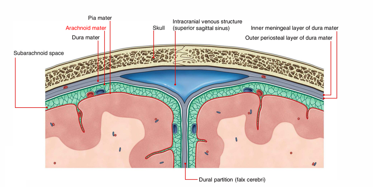
The arachnoid mater is a slim, avascular membrane which outlines, but is not conjoined to the inner surface of the dura mater and it is easily separable with the dura mater with the exception of the point where it is conjoined to the adventitia of the internal carotid as well as vertebral arteries where they move within the subarachnoid space.

Arachnoid Mater
- With the exception of the great longitudinal fissure in the middle of the cerebral hemispheres, it does not move within the sulci or fissures. Through a system of connective tissue which travels from the subarachnoid space referred to as arachnoid trabeculae, it is roughly connected together with the pia mater.
- The pituitary gland itself is not surrounded by it; however it spreads out towards the superior surface of the pituitary fossa. As a part of the optic nerve-sheath complex the arachnoid mater also leaves the cranium.
- Compared to the pia, with the exception of the longitudinal fissure between the two cerebral hemispheres, the arachnoid does not go into the grooves or fissures of the brain. Thin processes or trabeculae through its inner surface spread out downwards, then travel through the subarachnoid space, and end up being constant together with the pia mater.
- At the foramen magnum the arachnoid mater proceeds as the spinal arachnoid, which terminates at the level of second sacral vertebra. It has exactly the same form as the dural sac with the exception of the point where its arachnoid granulations perforate the dura mater and is closely related to the internal surface of the dura mater.
- A capillary space called the subdural space containing a film of fluid splits up the arachnoid mater from the dura mater. Cerebral veins on their route towards the dural venous sinuses travel through the subdural space. Surrounded by the arachnoid and pia mater, this creates a sliding plane where movement is achievable amongst the dura mater and brain.
Substructures of Arachnoid
Arachnoid villi
These originate from the surface of arachnoid and are fine finger-like projections. They eventually perforate dura in order to project within the dural venous sinuses, before that they move the dura before them. Specific mesothelial cells that convey the CSF towards the bloodstream surround them, as a result leading to the absorption of CSF.
Arachnoid granulations (Pacchionian Bodies)
With progression of age, pedunculated tufts called arachnoid granulations are created and the arachnoid villi increase in size. A general consideration is that the arachnoid granulations are the large clusters of arachnoid villi. Arachnoid granulations are involved with the absorption of CSF like arachnoid villi. They protrude within the venous lacunae of the superior sagittal sinus.

