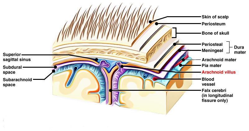
Arachnoid villi originate from the surface of arachnoid mater and are thin finger-like projections. Human arachnoid villi have four parts:
- Fibrous capsule
- Arachnoid cell layer
- Cap cell cluster
- Central core
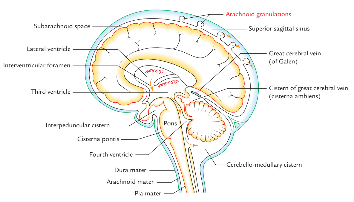
They eventually perforate dura in order to project within the dural venous sinuses, before that they move the dura before them. Specific mesothelial cells that transport the CSF towards the bloodstream surround them, as a result leading to the absorption of CSF. The CSF goes back toward the venous system via arachnoid villi. These project as clumps into the superior sagittal sinus, which is a dural venous sinus, and its lateral extensions, the lateral lacunae.

Arachnoid Villi
Boundaries
A thin fibrous capsule with an endothelial covering being reflected from the venous sinus lumen mainly covers the arachnoid cell layer surrounding the central core. These endothelial cells are joined by desmosomes and light junctions and have numerous micropinocytotic vesicles, microvilli, as well as some large vacuoles.
Cells of the adjacent dura and are composed of connective tissue fibers, the fibrous capsule are strengthened by a coating of electron-dense flattened cells with long projections same as dural border. Particularly at the apical part of the villus, the fibrous capsule is often very thin or insignificant. The arachnoid cell layer borders straight over the lumen of lateral lacuna or the sinus.
Arachnoid Granulations (Pacchionian Bodies)

Arachnoid Granulations
A general consideration is that the arachnoid granulations are the large clusters of arachnoid villi. Because they were first described by the Italian scientist Antonio Pacchioni in 1705, they are also called “Pacchionian granulations”. Arachnoid granulations are involved with the absorption of CSF like arachnoid villi. They protrude within the venous lacunae of the superior sagittal sinus. They are closely related not only to the absorption of CSF but also to the manifestation of meningiomas. With progression of age, pedunculated tufts called arachnoid granulations are created and the arachnoid villi increase in size.
While arachnoid villi are abundant and mainly protrude within the lateral lacunae of the superior sagittal sinus, arachnoid granulations are small in quantity and directly project within the inner table of the skull by means of perforation of dura mater or within the major venous sinus lumen.

 (74 votes, average: 3.95 out of 5)