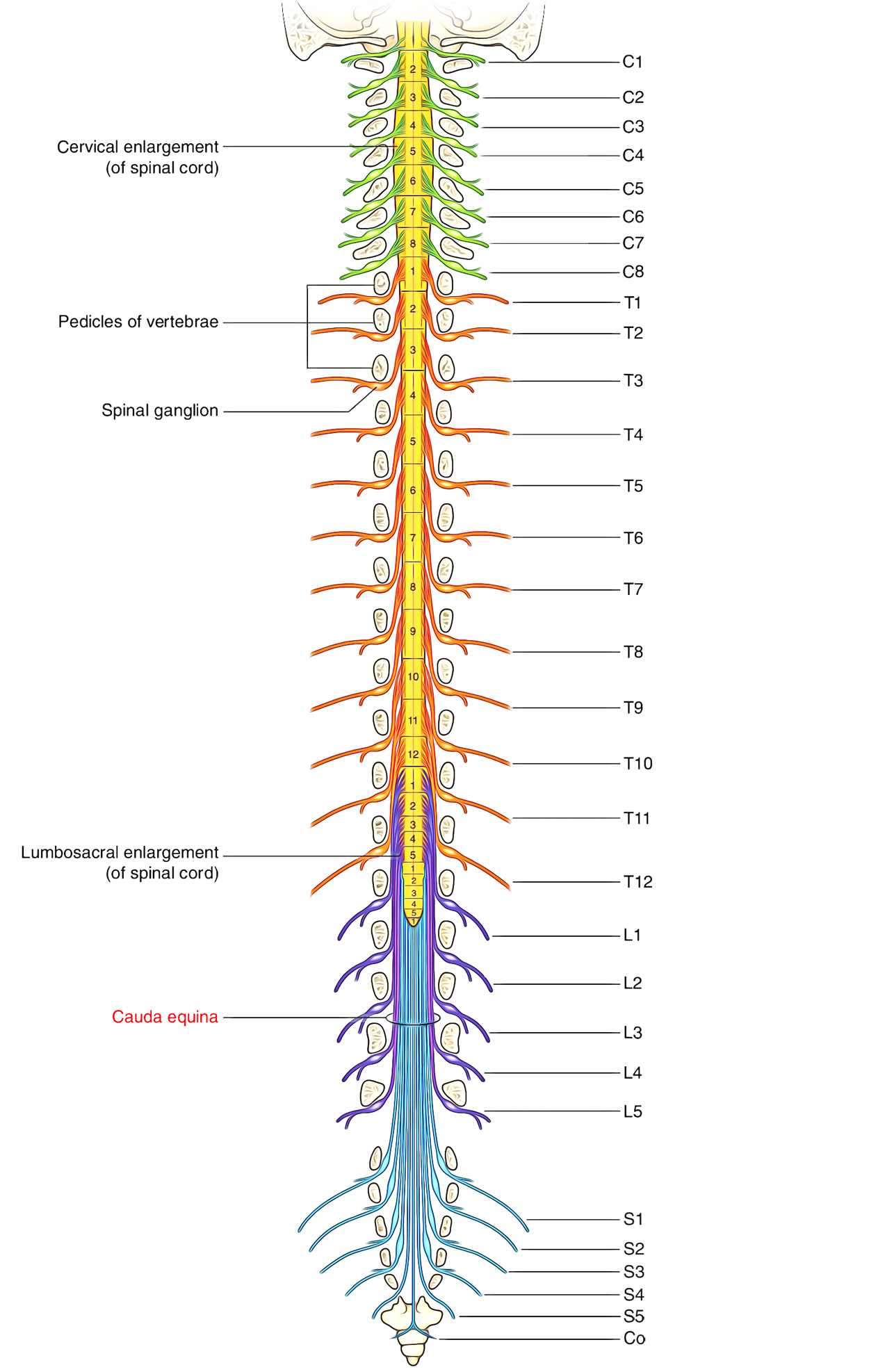
Cauda equina is the package of nerves comprised of the lumbar, sacral as well as coccygeal nerve roots coming from L2 to S5 creates cauda like a horse tail. Generally it starts at the level of L1/L2 disc space distal towards the conus medullaris. The spinal cord terminates at the lower border of the first lumbar vertebra where it blends to create the conus medullaris. The lumbar as well as sacral nerve roots which emerge from the lower portion of spinal cord create a bundle inside the spinal canal under the conus medullaris. This bundle of nerves is identical in appearance to a horse’s tail and is referred to as the cauda equina. Lumbar as well as sacral nerve roots exit behind the spinal canal, as well as send and collect nerve stimulus to and from the lower limbs along with pelvic organs.

Cauda Equina
Development
Spinal cord is located opposite S2 vertebral level at the time of the foetal growth, on day 22. It is located towards L5 level on day 70 as longitudinal progression of vertebrae out grows the length of spinal cord. On day 160 it is located opposite towards L2 which is its normal level at birth. Then distal neural tube degenerates and transforms into filum terminale. This remains connected to S2 vertebral body.
Artery Supply
Lower spinal cord along with the nerve roots of the cauda equina is supplied by various vessels going into the canal on one portion of upper lumbar roots. Accessories from iliolumbar along with lateral sacral divisions of internal iliac artery are the primary supply of this particular zone.
Clinical Significance
Cauda Equina Syndrome (CES) arises when the nerves inside the spinal canal have been injured. Consequently, the nerves providing the muscles of the legs, bladder, bowel, as well as genitals do not work efficiently. Patients encounter desensitization, loss of sensation along with pain in the legs, buttocks, and pelvic zone to altering degrees. The damage is usually irreversible.
Start
Partial and total cauda equina syndromes are common. The cauda equina generally starts at the level of the 1-1/2 disc space, distal towards the conus medullaris. This bundle of nerves is made up of the lumbar, sacral, as well as coccygeal nerve roots via 1.2 to C0. Early medical diagnosis and surgical treatment may greatly improve outcome. The cauda equina is more immune towards injury compared to the spinal cord.
Symptoms
- Generally, patients with approximately partially maintained sensation found with weak or flaccid lower extremities.
- Knee and ankle tugs are absent. Sensory loss usually is disproportional and may be radicular, instead of the sensory disabilities seen in the conus medullaris syndrome, which are more proportional.
- So-called saddle anesthesia is one of the most common sensory deficiencies.
- Saddle anesthesia signifies loss of sensation near the anus, genitals, perineum, buttocks, as well as posterior-superior thighs.
- Loss of bowel, bladder, and sexual functionality is also common.
- Urinary retention is one of the most common features of a cauda equina syndrome because in the absence of urinary retention there is only a 1 in 1000 possibility of acquiring a cauda equina syndrome.
- The cauda equina syndrome may be complete, though rarely. Sensory axons are a lot more resistant to damage compared to motor axons. The in-depth signs depend upon the level of the injury
Causes of Cauda Equina Syndrome
There are eight common reasons for cauda equina syndrome.
- Congenital Reasons.
- Arachnoiditis.
- Vascular.
- Trauma/Injury.
- Herniated disc.
- Spinal stenosis.
- Tumours (Neoplasms).
- Accidental medical reasons (Iatrogenic causes).

 (56 votes, average: 4.85 out of 5)