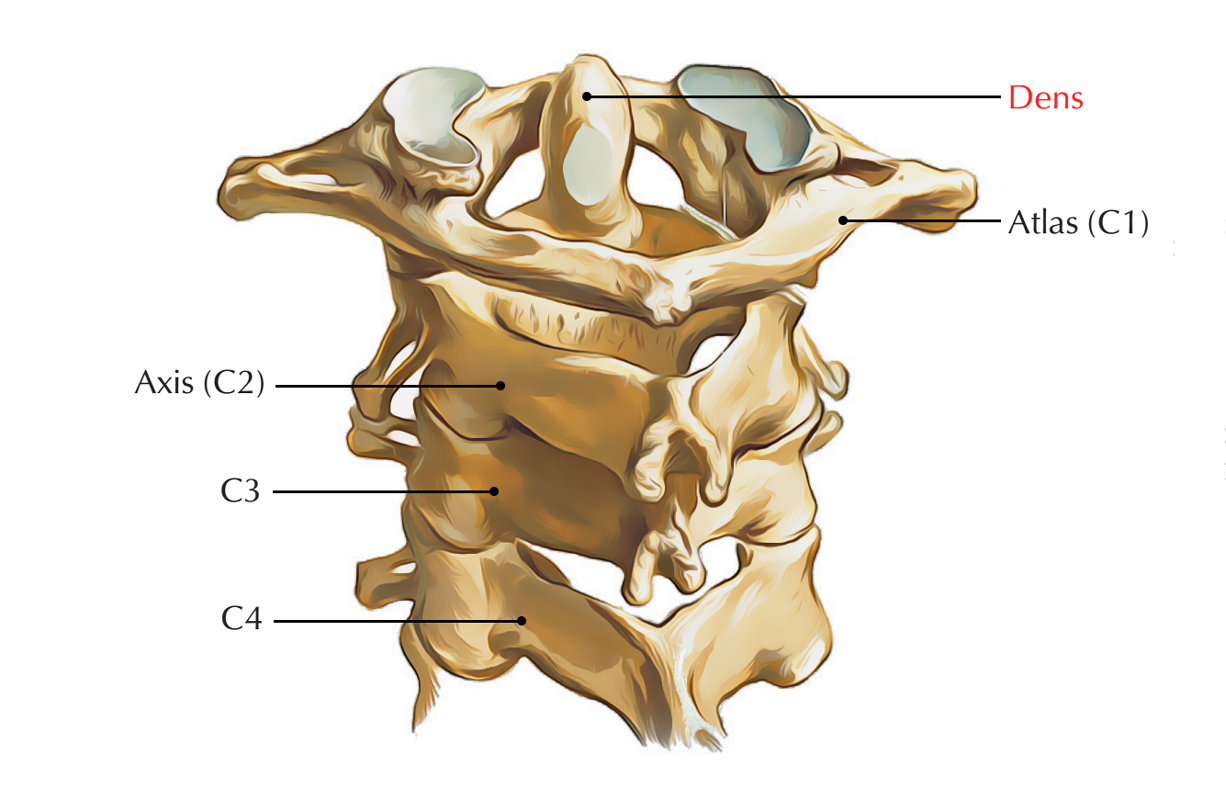
The dens is a protuberance (process or projection) of the C2 vertebra or axis which is also called the odontoid process. It is the most prominent feature of the c2 vertebra. It has a small contraction where it connects with the main body of the vertebra.
It is originally made up of an upward extension of the cartilaginous mass, inside the cartilaginous mass the lower part of the body is created.

Dens
Structure
Internal Structure
Compared to the body, the internal structure of the dens is more compressed. The odontoid process is the ascension of the atlas merged with the ascension of the axis.
- The inner ligaments are very strong and inhibit the rotation of the head.
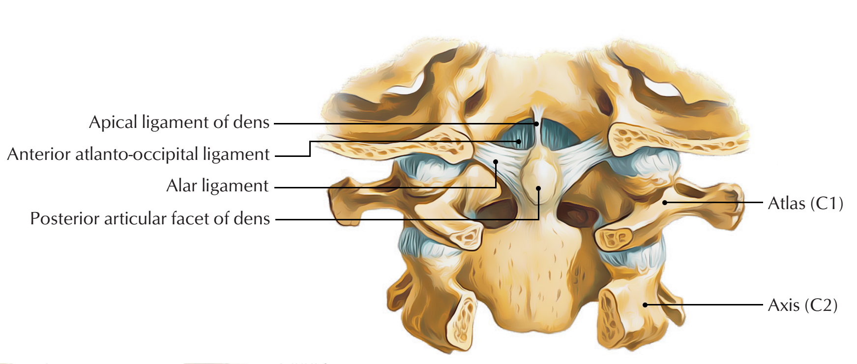
- The weak apical ligament is located anterior towards the upper longitudinal bone of the cruciform ligament and connects with the apex of the deltoid peg towards the anterior border of the foramen magnum.
- At its front, the process possesses an articular facet and together with the anterior arch of the atlas, it creates a portion of the joint and is a non-weight bearing joint.
The alar ligaments and the apical ligaments are attached towards the borders of the foramen magnum from the inclined upper edge of the odontoid process.

Dens: Structure
Apex
- The apex is sharp and the apical odontoid ligament connected to it.
- The process is a bit engorged under the apex and a rough depression is found for the connection of the alar ligament on either side of it.
Attachments
- A shallow groove for the transverse atlantal ligament which holds the process at its place is present posterior to the neck and often spreading towards its lateral surfaces.
- The occipital bone and the process linked by these ligaments.
- An oval or almost circular aspect for the creation of joint with anterior arch of the atlas is present on its anterior surface.
Development
The dens or odontoid process originally is made up of an upward extension of the cartilaginous mass, inside which the lower part of the body is created.
- Around the sixth month of fetal life, two centers make their appearance at the base of this process:
- They are laterally located and join before birth in order to create a conical bilobed mass deeply cleft above.
- A wedge-shaped portion of cartilage creates the gap in the middle of the sides of the cleft and the apex of the process.
- A cartilaginous disk divides the base of the process from the body.
Clinical Significance
Fractures
Based on the Anderson / D’Alonso system, fractures of the dens are categorized into three types:
- Type I: This is generally a stable type of fracture. It spreads out via the apex of the dens.
- Type II: For this region of the axis, it is the most frequently acquired fracture. It has a high rate of non-union form and is unsteady. It spreads out via the base of the dens.
- Type III: It can be either stable or unstable and surgery may be needed. It spreads out from the vertebral body of the axis.

 (52 votes, average: 4.77 out of 5)