The flexor digitorum superficialis (FDS) is the biggest muscle of the superficial group of muscles on the front of the forearm. It creates the intermediary muscle layer in between the superficial and deep groups of the forearm muscles.

Flexor Digitorum Superficialis
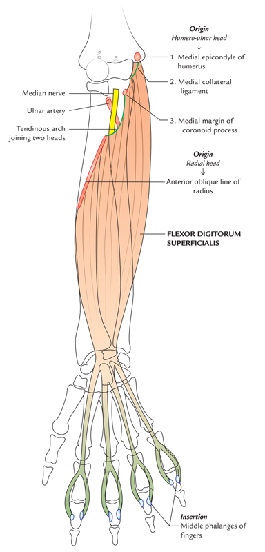
Origin
It emerges by two heads:
- humeroulnar head, via the medial epicondyle of humerus, superb tubercle on the medial margin of the coronoid process of ulna and medial (ulnar) collateral ligament of the elbow joint.
- Radial head, via the anterior oblique line of the radius, extending via the radial tuberosity to the attachment of pronator teres (upper half of the anterior border of radius).
Flexor Digitorum Superficialis: Origin and Insertion
Structure
- The flexor digitorum superficialis is a broad, relatively large muscle layer in the forearm, primarily deep however partially superficial.
- The superficial part comes to the surface at numerous locations, hut can just sometimes be seen directly, when the fist is clenched, in the long narrow periods in between the flexor carpi radialis, palmaris longus, and flexor carpi ulnaris.
- The subcutaneous tendon of the flexor digitorum superficialis to the ring finger is normally effortlessly seen, being located in between the tendons of the palmaris longus and flexor carpi ulnaris on the distal quarter of the forearm.
- When the hand is clenched within a fist and the wrist is somewhat bent it is most easily seen.
- The tendon of the flexor digitorum superficialis to the middle finger glide sideways (towards the ulna) out via under the tendon of the palmaris longus if the hand is then turned towards the ulnar side.
- The tendons of the flexor digitorum superficialis all push the similar plane as they move distally to their particular fingers in the palm.
- These tendons might be seen in relief on the palm, radiating via the wrist towards the fingers when the palmar fat is thin, and the fingers are partially bent against resistance.
Attachment
Middle phalanges of medial four fingers
The mode of attachment is as follows. The muscle divides into two layers: superficial and deep.
- The superficial layer creates two tendons, which are placed within middle phalanges of middle and ring fingers.
- The deep layer likewise forms two tendons, which are placed within middle phalanges index and little fingers.
Before attaching to each of the four tendons, divides opposite to the proximal phalanx, within medial and lateral slips, which are placed within the matching sides of the middle phalanx.
- The median nerve and ulnar artery move downwards deep to the fibrous arch/tendinous arch linking the humeroulnar and radial heads of flexor digitorum superficialis.
- The four tendons of flexor digitorum superficialis move deep to flexor retinaculum confined within a common synovial sheath, the ulnar bursa.
Actions
- Flexor digitorum superficialis bends the proximal interphalangeal (PIP) joints of the medial four digits.
- Operating more highly, it also assists in flexion of the proximal phalanges and wrist joint.
Nerve supply
The flexor digitorum superficialis is supplied by the median nerve.
Rate this Article: 
 (53 votes, average: 4.78 out of 5)
(53 votes, average: 4.78 out of 5)