The basic structure of human body is skeletal system. The entire body is built around the hard framework of Skeleton. At birth around 270 bones are present, by adulthood this total decreases to around 206 bones when bones merge together. About the age of 21 the bone density within the skeleton achieves its full density.
[skeleton]Divisions of Skeleton
The human skeleton can be distributed into the axial skeleton and the appendicular skeleton:
- The axial skeleton is created by the vertebral column, the rib cage, the skull and other associated bones.
- The appendicular skeleton, which is connected with the axial skeleton, is created by the shoulder girdle, the pelvic girdle as well as the bones of the upper and lower limbs.
Axial Skeleton
Skull
- Mandible
- Occipital bone, two temporal bones, two parietal bones, the sphenoid, ethmoid and frontal bones.
- Vomer, two nasal conchae, two nasal bones, two maxilla, the mandible, two palatine bones, two zygomatic bones, and two lacrimal bones.
- The occipital, parietal, frontal, in the neurocranium, and the nasal, lacrimal, and vomer
Vertebrae
Specific vertebrae are entitled corresponding to their area as well as location. The vertebrae are from highest to lowest:
- Cervical spine: 7 vertebrae (C1–C7)
- Thoracic spine: 12 vertebrae (T1–T12)
- Lumbar spine: 5 vertebrae (L1–L5)
- Sacrum: 5 (fused) vertebrae (S1–S5)
- Coccyx: 4 (3–5) (fused) vertebrae (Tailbone)
Rib Cage
It contains 24 ribs that are made of 12 pairs, 12 ribs each side.
Appendicular Skeleton
The appendicular skeleton is split into six major regions:
- Pectoral girdles (4 bones) – Left and right clavicle (2) and scapula (2).
- Arms and forearms (6 bones) – Left and right humerus (2) (arm), ulna (2) and radius (2) (forearm).
- Hands (54 bones) – Left and right carpals (16) (wrist), metacarpals (10), proximal phalanges (10), intermediate phalanges (8) and distal phalanges (10).
- Pelvis (2 bones) – Left and right hip bone (2).
- Thighs and legs (8 bones) – Left and right femur (2) (thigh), patella (2) (knee), tibia (2) and fibula (2) (leg).
- Feet and ankles (52 bones) – Left and right tarsals (14) (ankle), metatarsals (10), proximal phalanges (10), intermediate phalanges (8) and distal phalanges (10).
Functions of the Skeletal System
The skeletal system performs several basic functions:
- Support: The skeleton acts such as the structural framework for the body by supporting soft tissues as well as giving connection points for the tendons of majority of skeletal muscles.
- Protection: The skeleton guards the majority of vital interior organs from injury.
- Assistance in movement: Mainly skeletal muscles connect to bones; as soon as they contract, they produce movement by pulling on the bones.
- Mineral homeostasis (storage and release): Bone tissue stores numerous minerals, particularly calcium and phosphorus, which add to the strength of bone. Bone tissue stocks around 99% of the body’s calcium. Bone discharges minerals within the blood for sustaining vital mineral equilibrium (homeostasis) as well as for supplying the minerals towards other parts of the body.
- Blood cell production: Inside specific bones, a connective tissue known as red bone marrow creates red blood cells, white blood cells, along with platelets, in a process named hemopoiesis. Red bone marrow contains maturing blood cells, adipocytes, fibroblasts, as well as macrophages inside a linkage of reticular fibers. It exists in growing bones of the fetus as well as in some adult bones, like the pelvic bones, ribs, sternum, vertebrae, skull, and tips of the humerus along with femur. In a baby, total bone marrow is red and is engaged in hemopoiesis. With growing age, the majority of the bone marrow turns from red to yellow.
- Triglyceride storage: Yellow bone marrow contains mostly of adipose cells, which stock triglycerides. The stockpiled triglycerides are a latent chemical energy backup.
Components of the Skeleton
Fibrous and mineralized connective tissues create the skeleton, which gives it firmness and flexibility. It consists of:
Bone: It is made up of collagen and calcium phosphate, a mineral crystal, and is a type of mineralized connective tissue. Bones get their hardness from Calcium phosphate. Bone tissue can be compact or spongy. Bones give structural support and protection for body organs.
Cartilage: it is composed of compactly filled collagenous fibers inside a rubbery gelatinous substance called chondrin and is a type of fibrous connective tissue. Cartilage gives flexible support for certain structures in adult humans including the nose, trachea, and ears.
Tendon: It is bonded to bone and connects bone to bone and is a fibrous band of connective tissue.
Ligament: a fibrous band of connective tissue that joins bones and other connective tissues together at joints.
Joint: a location where two or more bones or other skeletal components are joined together.
Bone
Bone creates the most of the skeleton and is a calcified, living, connective tissue. It contains an intercellular calcified matrix, which also consists of collagen fibers, as well as numerous varieties of cells within the substance. Bones function as:
- supportive structures for the body
- protectors of vital organs
- reservoirs of calcium and phosphorus
- levers on which muscles act to produce movement
- Containers for blood-producing cells.

Human Skeletal System: Bone
Types
Two varieties of bone are seen. Classification according to the structure:
- Compact bone is dense bone that creates the outer covering of all bones as well as surrounds spongy bone.
- Spongy bone consists of spicules of bone surrounding cavities having blood-making cells (marrow).
Classification of the bones according to the shape:
- Long bones are tubular (e.g., humerus in upper limb; femur in lower limb).
- Short bones are cuboidal (e.g., bones of the wrist and ankle).
- Flat bones consist of two compact bone plates separated by spongy bone (e.g., skull).
- Irregular bones are bones with various shapes (e.g. bones of the face).
- Sesamoid bones are round or oval bones that develop in tendons.
Cartilage
Cartilage is an avascular type of connective tissue. Which contains extracellular fibers entrenched within a matrix that contains cells localized in small cavities. The quantity and type of extracellular fibers inside the matrix alters dependent on the type of cartilage. The functions of cartilage are:
- Support soft tissues.
- Provide a smooth, sliding surface for bone articulations at joints.
- Enable the development and growth of long bones.

Human Skeletal System: Cartilage
Types
There are three types of cartilage:
- Hyaline: most common type in which the matrix contains a moderate amount of collagen fibers (e.g., articular surfaces of bones).
- Elastic: matrix contains collagen fibers along with a large number of elastic fibers (e.g., external ear).
- Fibrocartilage: matrix contains a limited number of cells and ground substance amidst a substantial amount of collagen fibers (e.g., intervertebral discs).
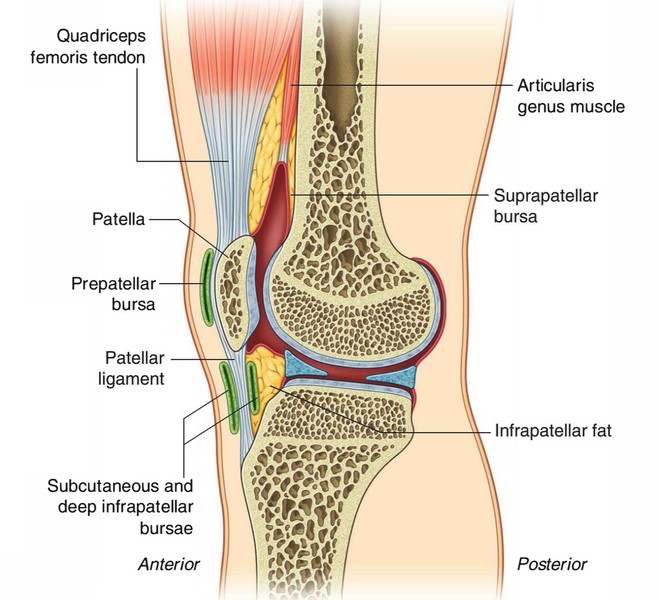
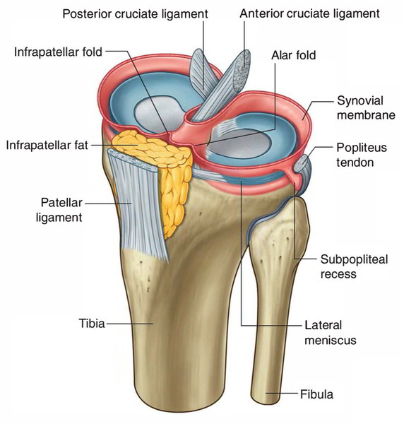
Joints
The places where two skeletal elements merge are known as joints. The two common categories of joints are those in which:
- The skeletal elements are separated by a void (i.e., synovial joints).
- There is no void and then the components are connected via a connective tissue (i.e., solid joints).

Human Skeletal System: Joint
Blood vessels which traverse a joint as well as nerves which innervate muscles working over a joint usually give articular branches towards that particular joint.
Tendons
A tendon usually links muscle to bone and is able to endure strain, and is a tough strap of fibrous connective tissue. Tendons and ligaments both are created by collagen. Ligaments join one bone to another bone, while tendons connect muscle to bone.
Ligaments
A strap of dense normal connective tissue packs made of collagenous fibers, by bundles secured via condensed rough connective tissue coverings. Ligaments join bones to other bones to form joints; however tendons join bone to muscle. Certain ligaments restrict the movement of articulations, or prevent certain activities completely.

 (49 votes, average: 4.88 out of 5)