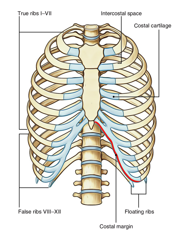
The ribs stretches posteriorly from thoracic vertebrae to the anterior lateral edges of the sternum. They are ribbon like, elastic bony arches and flat in shape. Coastal cartilages are joined to the anterior ends. The costal cartilage and ribs together makes the costa. Also the large part of the thoracic skeleton is created by the ribs and their costal cartilages.
Ribs
Number
Normally there are 12 pairs of ribs (but incidence of accessory cervical or lumbar rib may grow them to 13 pairs or absence of 12th rib may reduce them to 11 pairs).
Arrangement and General Outline
- The ribs are arranged 1 below the other and the gaps between the adjacent ribs are termed intercostals spaces.
- The length of ribs increases from 1st to 7th rib and after that slowly falls; therefore, seventh rib is the longest rib.
- The transverse diameter of thorax increases progressively from 1st to 8th rib, thus 8th rib has the best lateral projection.
- The ribs are arranged obliquely, i.e., their anterior ends be located at lower level than their posterior ends.
- The obliquity of ribs rises progressively from 1st to 9th rib, for this reason 9th rib is most obliquely set.
- The width of ribs slowly reduced from above downward.
The anterior ends of first 7 ribs are joined to the sternum via their costal cartilages. The cartilages at the anterior ends of 8th, 9th, and 10th ribs are joined to the next higher cartilage. The anterior ends of 11th and 12th ribs are free and hence named floating ribs.
- The 10th rib generally have free anterior ends in Japanese
- First rib slopes downwards along its whole extent
- The middle of every costal arch (being composed of a rib and its costal cartilage) with the exception of the very first rib is located at a lower level when compared to a straight line joining the 2 ends of the costa
Categorization
According to Features
- Typical ribs: 3rd-9th.
- Atypical ribs: 1st, 2nd, 10th, 11th, and 12th.
The normal ribs have same general features, on the other hand the atypical ribs have special features and thus can be discerned from the rest of the ribs.
According to Relationship with the Sternum
- True ribs: lst-7th (i.e., upper 7 ribs).
- Bogus ribs: 8th-12th (i.e., lower 5 ribs).
True ribs articulate with the sternum anteriorly, on the other hand false ribs don’t articulate with the sternum anteriorly.
According to Articulation
- Vertebrosternal ribs: lst-7th.
- Vertebrochondral ribs: 8th-10th.
- Vertebral (floating) ribs: 11th and 12th.
The vertebrosternal ribs joint posteriorly with vertebrae and anteriorly with the sternum.
The vertebrochondral ribs joint posteriorly with vertebrae and anteriorly their cartilages join the cartilage of the higher rib. The vertebral or floating ribs joint posteriorly with the vertebrae but their anterior ends are free.
Typical Ribs
Ribs: Typical Ribs
Parts
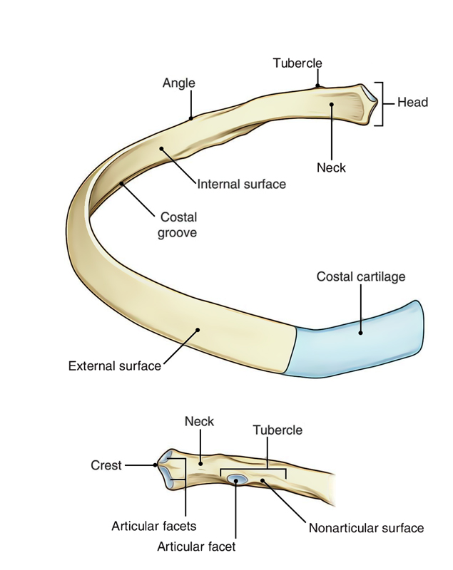
Every rib has 3 parts: (a) anterior end, (b) posterior end, and (c) shaft.
- The anterior end bears a concave depression.
- The posterior end is composed of head, neck, and tubercle.
- The shaft is the longest part and goes between anterior and posterior ends. It’s flattened and has inner and outer surfaces and upper and lower edges. It’s arch with convexity directed outwards and bears a costal groove on its inner surface near the lower border. 5 centimeters far from tubercle, it suddenly changes its direction, this is termed angle of the rib.
Side Conclusion and Anatomical Position
The side of the rib can be figured out by holding it in this type of style that its posterior end having head, neck, and tubercle is directed posteriorly, its concavity faces medially and its sharp border is directed inferiorly.
In an anatomical position, the posterior end is higher and nearer the median plane in relation to the anterior end.
Features and Attachments
Anterior (Costal) End
It carries a small cup shaped depression, which joins the corresponding costal cartilage to create a primary cartilaginous costochondral joint.
Posterior End
It presents head, neck, and tubercle.
Head
It’s 2 articular facets: lower and upper.
- The lower bigger facet articulates with the body of numerically corresponding vertebra
- The upper smaller facet articulates with the next higher vertebra
The crest separating the 2 articular facets is located opposite the intervertebral disc.
Neck
- It is located in front of the transverse process of the corresponding vertebra.
- it’s 2 edges – superior and inferior, and 2 surfaces– anterior and posterior.
- The upper border is sharp crest like, on the other hand the lower border is rounded.
- The posterior surface is rough and pierced by foramina. Tubercle.
- It’s situated on the outer outermost layer of the rib in the junction of neck and shaft.
- It’s divided into 2 parts – medial articular part and lateral non-articular part. The articular part bears a small oval facet, which articulates with all the transverse process of corresponding vertebra. The non-articular part is rough and gives connection to ligaments.
Shaft
- It’s thin and flattened.
- It presents 2 surfaces – outer and inner, 2 edges – upper and lower, and 2 angles – posterior and anterior.
Edges
Superior Border
The superior border is thick and rounded, and presents outer and inner lips:
- The outer lip gives connection to the external intercostal muscles.
- The inner lip gives connection to the internal intercostal and intercostalis intimus muscles.
Lower Border
The lower border is sharp and creates the lower border of the costal groove, and gives origin to the external intercostals muscle.
Surfaces
Outer Surface
- It’s smooth and convex, and presents 2 angles– posterior and anterior.
- The posterior angle (typically referred to as only angle) is marked by an oblique ridge.
- The anterior angle is marked by an indistinct oblique line.
Inner Surface
It’s smooth and concave. It presents a costal groove near its lower border. The costal groove becomes unrecognizable in the anterior part.
- The costal groove lodges intercostal nerve and vessels from above downwards, as follows (Mnemonic: VAN):.
- Intercostal Vein.
- Intercostal Artery.
- Intercostal Nerve.
- The internal intercostal muscle is connected to the floor of the groove (interceding between intercostal nerves and vessels, and bone).
- The intercostalis intimus is connected to the upper border of the costal groove.
3 Characteristic Features of Atypical Rib:
- It’s curved along its whole extent
- It’s angulated, i.e. presents 2 curves 1 5 cm in front of tubercle and 1 2 cm behind the anterior end
- It’s twisted, in order that both ends of the rib can not contact the same horizontal plane
(Mnemonic: CAT – Curve, Angle, and Twisted)
Atypical Ribs
First Rib
Distinguishing Features
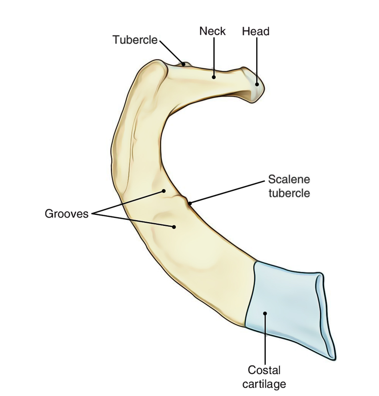
- It’s shortest, widest, and most intensely arch.
- Its shaft is flattened above downwards so that it’s upper and lower surfaces, and outer and inner edges.
- Its head is small, rounded, and bears a single circular articular facet to joint with the side of first thoracic vertebra.
- Its angle and tubercle coincide.
- it’s no costal groove on its inner surface.
- Its neck is rounded and elongated. It’s pointed upwards, backwards and laterally.
- Its anterior end is bigger and thicker.
Ribs: First Rib
The shaft of the very first rib slopes obliquely downwards and forwards to its sternal end. It’s because of this obliquity the pulmonary and pleural apices projects into the root of the neck.
Side Decision
Side of the very first rib can be figured out by supporting the rib in this manner that:
- Its bigger end is directed anteriorly and its smaller end is directed posteriorly
- The surface of its shaft having 2 grooves divided by a ridge is directed superiorly
- Its concave border is directed inwards and its convex border is directed outwards
Trick For Pupils For Side Determination Of First Rib:
Keep the rib on the table top contemplating its position in your body. Now notice the rib belongs to the side on which it is both ends touch the surface. If the rib is set on the incorrect side, then only its anterior end will be contacting the surface.
Features And Attachments
Inner Border
- It presents a scalene tubercle about its middle. Tubercle and adjoining part of the upper surface gives connection to the scalenus anterior muscle
- It gives connection to the Sibson’s fascia (suprapleural membrane)
Outer Border
It gives origin to the first digitation of serratus anterior about its middle, just behind the groove for the subclavian artery.
Superior (Upper) Surface
- It’s crossed obliquely by 2 shallow grooves (anterior and posterior) divided by a little ridge. The ridge is constant with the scalene tubercle. The anterior groove lodges subclavian vein, while posterior groove lodges the subclavian artery and lower trunk of the brachial plexus
- The area behind posterior groove up to the costal tubercle gives connection to the scalenus medius muscle
- The area in front anterior groove and near the anterior end gives connection to subclavius muscle (anteriorly) and costoclavicular ligament (posteriorly)
Lower Surface
It’s related to the costal pleura.
Neck
- It’s elongated and pointed upwards, backwards, and laterally
- The subsequent structures create anterior relationships of neck from medial to lateral side
- Sympathetic chain
- First posterior intercostal Vein
- Superior intercostal Artery
- Ventral ramus of first thoracic Nerve
Memory apparatus: Chain pulling the VAN
Second Rib
Ribs: Second Rib
Distinguishing Features and Attachments
- Its length is twice that of the next rib
- Its shaft is sharply/ tremendously bent
- Its shaft isn’t twisted; thus both the ends of rib touch the table top when put on it
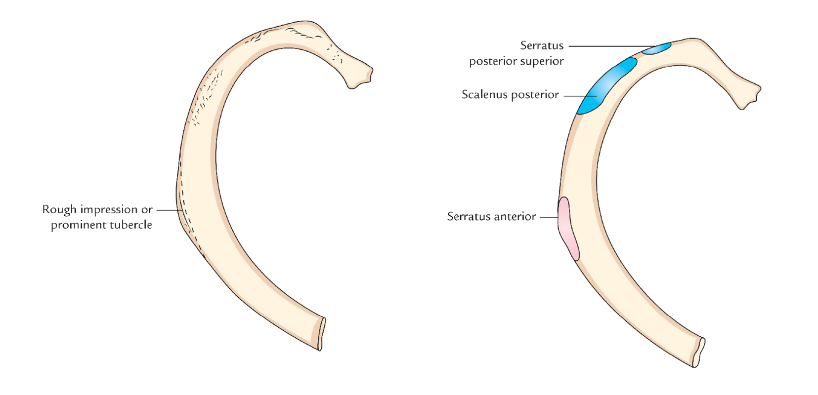
- Near its middle, the outer convex surface of shaft presents a rough impression or notable tubercle, which gives connection to the serratus anterior muscle (lower part of first and whole of 2nd digitation)
- The outer outermost layer of the shaft is pointed outwards and upwards, while inner outermost layer of the shaft is pointed inwards and downwards
- Posterior part of internal surface presents a short costal groove
- The upper border and adjoining part of upper surface supply connection to the scalenus posterior and serratus posterior superior muscles
Tenth Rib
Distinguishing Features
- It’s single articular facet on its head, which articulates with the body of corresponding thoracic vertebra
- It’s somewhat shorter in relation to the conventional rib
Eleventh Rib
Distinguishing Features
- It’s single large, articular facet on its head.
- It’s no neck and no tubercle.
- Its anterior end is pointed and tipped with cartilage.
- It’s little angle and a shallow costal groove.
- Its inner surface is pointed upwards and inwards.
- It’s a little angle.
Twelfth Rib
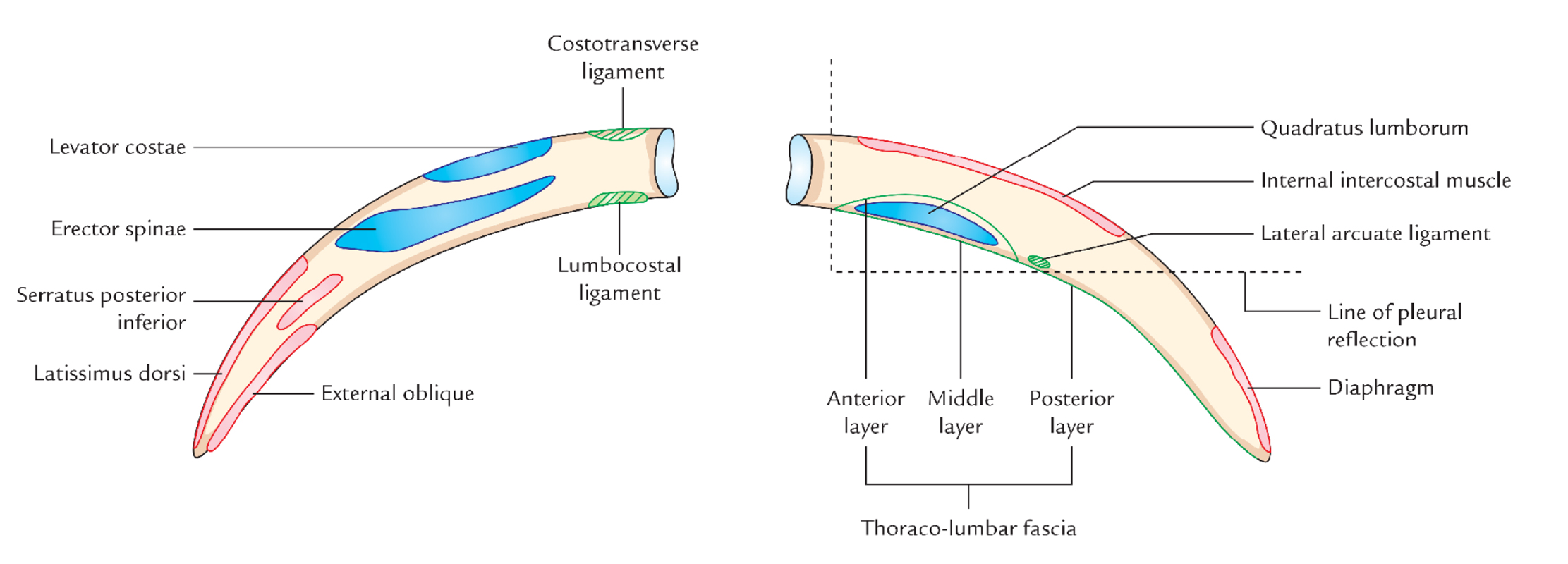
Ribs: Twelfth Rib
Distinguishing Features
The 12th rib has identical features as the 11th with the exception of that:
- It has no angle.
- It has no costal groove.
- It is considerably shorter than 11th.
Side Conclusion
The side of 12th rib can be figured out by keeping the rib in this style that:
- Its pointed anterior end is directed anterolaterally and its wider end posteromedially.
- Its somewhat concave surface faces inwards and upwards.
- Its sharper border is directed inferiorly.
Attachments
Outer Surface
- Near the tip, it gives connection to the latissimus dorsi muscle above and external oblique muscle below.
- Its medial half gives connection to the erector spinae muscle.
Inner Surface
- An oblique line crossing the middle of the surface indicates the line of pleural reflection.
- Lower part of medial half gives connection to the quadratus lumborum muscle.
- Middle two fourth near the upper border gives connection to the internal intercostal muscle.
- Upper part of sidelong quarter near the tip gives connection to the diaphragm.
Lower Border
- Close to head it gives connection to the lumbocostal ligament, which stretches from transverse process of LI vertebra.
- Lateral arcuate ligament is connected to it just lateral to the quadratus lumborum.
Upper Border
The upper border gives connection to the external and internal intercostal muscles.
Ossification
All the ribs ossify by 4 centres with the exception of 1st, 11th, and 12th ossify:
- 1 primary centre for shaft.
- 3 secondary centres: 1 for head, 1 for articular part of tubercle and 1 for non-articular part of the tubercle.
- First rib ossifies by 3 facilities: 1 primary center for shaft and 2 secondary facilities – 1 for head and 1 for tubercle.
- Eleventh and 12th ribs ossify by 2 facilities every: 1 primary centre for the shaft and 1 secondary centre for the head.
- Primary centres of all the ribs appear at the 8th week of IUL
- Secondary centres of all the ribs appear at puberty.
- Fusion in all the ribs happens at the age of 20 years.
Clinical Relevance
Cervical Rib
The costal component of the C7 vertebra may elongate to create a cervical rib in about 5% people. The state might be unilateral or bilateral. It takes place more commonly unilaterally and somewhat more frequently on the right side. The cervical rib might have a blind tip or the tip might be joined to the 1st rib by fibrous group or cartilage or bone. It might compress the lower trunk of brachial plexus and subclavian artery. The compaction generates: (a) pain along the medial side of forearm and hand and (b) interference in the circulation of the upper limb.
Lumbar Rib (Gorilla Rib)
It grows from the costal component of L1 vertebra. Its prevalence is much more common than the cervical rib, but stays undiagnosed as it normally doesn’t cause symptoms. It might be mistaken with the fracture of transverse process of L1 vertebra.
Fracture of Rib
Generally the middle ribs are included in the fracture. The rib normally fractures at its angle (posterior angle) as it’s the weakest point.
Flail Chest (Range-In-Chest)
When ribs are fractured at 2 sites (example, anteriorly as well as at an angle), the flail chest happens. The flail sections of ribs are sucked in during inspiration and pushed out during expiration resulting in a clinical condition referred to as paradoxical respiration.
Fracture of ribs is uncommon in kids as the ribs are elastic in them.
First 2 ribs (1st and 2nd ribs) are shielded by clavicle and last 2 ribs (11th and 12th) are mobile (floating), therefore they’re scarcely injured.

 (62 votes, average: 4.45 out of 5)