
Midbrain
- It includes the nuclei of the 3rd (oculomotor), 4th (trochlear) and 5th (trigeminal) cranial nerves. The midbrain is the smallest section of the brainstem and is situated just above the pons.
- It is traversed by the cerebral aqueduct. The part dorsal to the aqueduct is termed the tectum and is composed of 4 surface levels-the corpora quadrigemina (consisting of 2 superior and 2 inferior colliculi). The superior and inferior colliculi are concerned with reflexes calling for visual and auditory senses and get visual and auditory nerve impulses, respectively.
- The cerebral peduncles are split into left and right halves and are ventral to the aqueduct part. Every cerebral peduncle is composed of central part, the tegmentum, which is split ventrally from the crus cerebri by a mass of pigmented grey matter, the substantia nigra.
- The crus cerebri continues ipsilaterally with the internal capsule above and includes descending fibres from the cerebral cortex to the brainstem and spinal cord. The space between both crura is referred to as the interpeduncular fossa.
- The tegmentum mainly includes ascending tracts from the spinal cord to the thalamus and includes 2 large cigar shaped nuclei referred to as red nuclei. In cross section, the reddish nuclei appear as oval masses of the pinkish grey matter.
- The reddish nuclei help in unconscious management and coordination of motor tasks.
External Features
Features on the Ventral Aspect
- The ventral surface of the midbrain presents 2 crura cerebri which come from the cerebral hemispheres and converge downward to goes into the pons creating the posterolateral borders of the interpeduncular fossa.
- The superficial surface of the crus cerebri is elegantly corrugated by the inherent longitudinal fibres.
- The oculomotor nerve comes on either side from a groove on the medial side of the crus cerebri.
Features on the Dorsal Aspect
- The dorsal outermost layer of the midbrain presents 4 rounded elevations: 2 superior and 2 inferior colliculi (or corpora quadrigemina). The colliculi are divided from every other by a cruciform sulcus. The vertical limb of sulcus when followed above creates a surface depression which lodges the pineal body and when followed below, it becomes constant with the frenulum veli (a median ridge on the dorsal surface of superior medullary velum).
- The trochlear nerves come 1 on every side of the upper part of frenulum veli after decussation in the superior medullary velum. Thick ridges of white matter going from the lateral side of every colliculus make up their brachia.
- The superior brachium attaches the superior colliculus to the lateral geniculate body and the optic tract and is composed of optic tract fibres. The inferior brachium attaches the inferior colliculus to the medial geniculate body and is created from auditory fibres.
Internal Structure
- The internal structure of the midbrain is studied conveniently by analyzing its transverse sections. The transverse section of the midbrain reveals a miniature canal, termed cerebral aqueduct. A coronal plane going through the aqueduct splits the midbrain into 2 parts: a small posterior part and a large anterior part.
- The small posterior part is referred to as tectum and is composed of 4 colliculi. The large anterior part is split into 2 equivalent left and right halves, the cerebral peduncles by a vertical plane. Every cerebral peduncle is further subdivided into 3 parts; from dorsal to ventral, these are: (a) tegmentum, (b) substantia nigra and (c) crus cerebri.
- The structures in the tectum and tegmentum deviate at different levels of the midbrain, but those of the crus cerebri and substantia nigra are almost the same throughout the length of the midbrain.
Crus Cerebri (Basis Pedunculi)
The crus cerebri is the part of the cerebral peduncle situated anterolateral to substantia nigra. It includes essential descending tracts which attach the cerebral cortex to the anterior horn cells of the spinal cord, cranial nerve nuclei and pontine nuclei.
Substantia Nigra
- The substantia nigra is a curved (crescent shaped) pigmented band of grey matter (thicker medially than laterally) situated between tegmentum and crus cerebri. Its concavity is smooth and directed toward the tegmentum.
- The substantia nigra is created from intensely pigmented nerve cells which include melanin (a polymerized create of dopamine) and iron.
- The structure of tectum and tegmentum differ at different levels; for that reason, it’s again best studied by analyzing the T.S. of the midbrain at the subsequent 2 levels: (a) at the level of the inferior colliculi and (b) at the level of superior colliculi.
TS of Midbrain the Level of Inferior Colliculi
The grey and white matter at this level presents these features: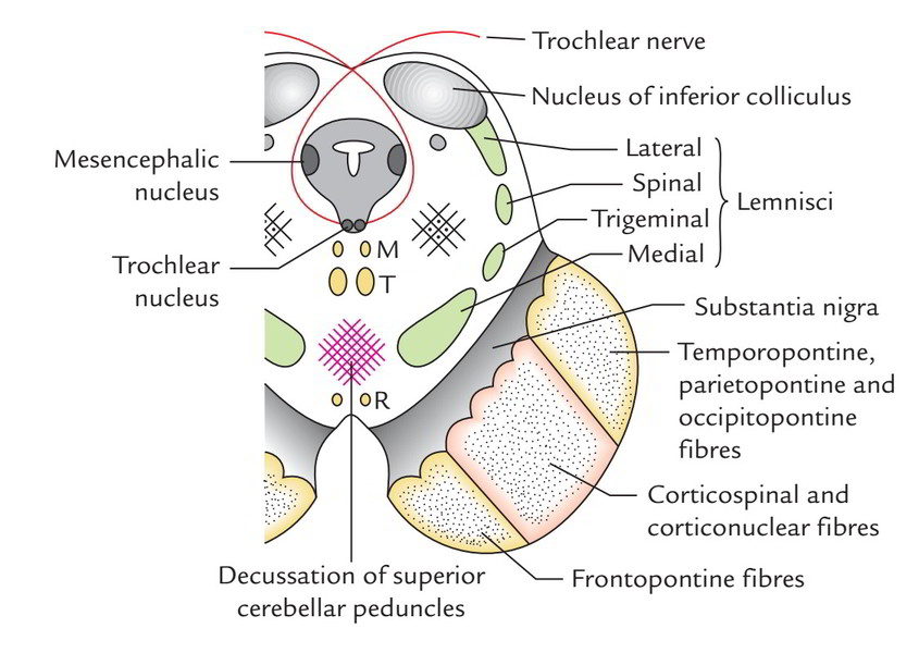
Grey Matter
- The central grey matter (grey matter around the cerebral aqueduct) includes 2 nuclei: (a) the nucleus of thetrochlear nerve and (b) the mesencephalic nucleus of the trigeminal nerve.
- The trochlear nerve nucleus is situated near the median plane just posterior to the MLF. The appearing fibres of the trochlear nerve pass laterally and posteriorly around the central grey matter and leave the midbrain just below the inferior colliculi. The fibres of the trochlear nerve now decussate in the superior medullary vellum before emerging on the surface.
- The mesencephalic nucleus of the trigeminal nerve is located in the lateral border of the central grey matter.
- An ovoid mass of grey matter underneath the inferior colliculus creates the nucleus of inferior colliculus.
White Matter
- The decussation of the superior cerebellar pedunclesoccupies the central part of the tegmentum and creates the most significant characteristic in the lower part of the middle-brain.
- The lemnisci are ordered in the creation of a curved streamlined band of white fibres in the ventrolateral part of the tegmentum, lateral to cerebellar decussation and dorsal to the substantia nigra. From medial to lateral side, these are: medial lemniscus, trigeminal lemniscus, spinal lemniscus and lateral lemniscus.
- The MLF is located on the side of the median plane ventral to the trochlear nerve nucleus.
TS of Midbrain at the Level of Superior Colliculi
Grey Matter
- The central grey matter in every half includes 2 nuclei: the oculomotor nerve nucleus and the mesencephalic nucleus.
- The oculomotor nucleus is located in the ventromedial part. The nuclei of 2 sides fuse together creating one complex having a triangular outline. The emerging fibres of oculomotor nerve pass ventrally via the tegmentum, intersecting red nucleus and medial part of the substantia nigra and emerge in the posterior part of the interpeduncular fossa via the sulcus on the medial aspect of crus cerebri.
- The mesencephalic nucleus takes up exactly the same position as in the lower part of the midbrain.
- The nucleus of superior colliculus is composed of cells that are affected in general light reflexes.
- Pretectal nucleus is a small group of nerve cells and is located deep to the superolateral part of the superior colliculus.
- Reddish nucleus is a cigar shaped mass of grey matter which seems ovoid in cross section. It’s about 0.5 cm in diameter. It’s situated in the tegmentum, ventral to the 3rd nerve nucleus and dorsomedial to the substantia nigra. It’s a significant nucleus of the extrapyramidal system.
White Matter
- Decussation of fibres (tectospinal and tectobulbar tracts) originating from superior colliculi creating dorsal tegmental decussation (of Meynert).
- Decussation of fibres (rubrospinal tracts) appearing from reddish nuclei creating ventral tegmental decussation (of Forel).
- Medial longitudinal fasciculus being located ventrolateral to the oculomotor nucleus.
- Tegmentum at this level also including the same lemnisci (i.e., medial, trigeminal and spinal) as those at the level of inferior colliculus with the exception of the lateral lemniscus.
- Emerging fibres of the oculomotor nerve.
Medial Longitudinal Fasciculus
- Medial longitudinal fasciculus is a greatly myelinated composite tract seen in the paramedian plane of the brainstem.
- The fibres of MLF interconnect the nuclei of the 3rd, 4th, 6th and 8th cranial nerves and the spinal nucleus of the accessory nerve.
- The main function of MLF is to organize the movements of eyes, head and neck in response to stimulation of the vestibulo-cochlear nerve.
Arterial Supply
The midbrain is supplied by the following arteries:
- Basilar artery via its posterior cerebral and superior cerebellar arteries. Basilar artery also supplies the midbrain via direct branches.
- Branches of posterior communication and anterior choroidal arteries.
Clinical Significance
Weber’s Syndrome
This syndrome is generated by a vascular lesion in the basal region of the cerebral peduncle as a result of occlusion of a branch of the posterior cerebral artery. This lesion requires the oculomotor nerve and the crus cerebri.
The characteristic features of the syndrome are as follows:
- Ipsilateral lateral squint, because of engagement of the 3rd cranial nerve.
- Contralateral hemiplegia, because of participation of corticospinal tract in the crus cerebri.
Benedict’s Syndrome
It takes place because of the vascular ischemia of the tegmentum of the midbrain, affecting medial lemniscus, spinal lemniscus, red nucleus, superior cerebellar peduncle and fibres of the oculomotor nerve. It’s defined by these signs and symptoms:
- Ipsilateral lateral squint and ptosis, as a result of participation of oculomotor nerve fibres.
- Contralateral loss of pain and temperature sense, because of engagement of trigeminal and spinal lemnisci.
- Contralateral loss of tactile, muscle, joint and shaking perception, as a result of participation of medial lemniscus.
- Contralateral tremors and involuntary movements in the limbs, because of participation of the red nucleus and fibres of superior cerebellar peduncle entering into it.
Perinaud’s Syndrome
It results from a lesion of the superior colliculi which happens when this area becomes compressed by the tumors of the pineal gland. It is defined by the loss of upward gaze without impacting the other eye movements.

 (60 votes, average: 4.80 out of 5)