The parietal bone creates the major portion of the vault of the skull and is a curved plate of bone. It’s quadrilateral in shape.
Parts
The parietal bone has:
- 2 surfaces: external and internal.
- 4 edges: superior (sagittal), inferior, anterior and posterior.
- 4 angles: frontal, sphenoidal, occipital and mastoid.
Surfaces
The 2 surfaces of the parietal bone are as follows:
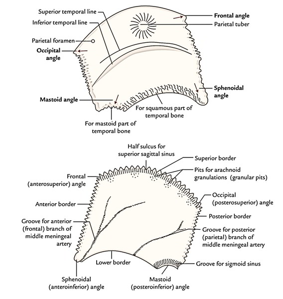
External surface: It’s most notable attribute of the surface that it is smooth and convex and is located near its center an elevation named parietal eminence or parietal tuberosity or parietal tuber. There are just two curved lines referred to as superior and inferior temporal lines, below the parietal tuberosity. The parietal foramen is situated near the posterior part of the superior border.
Internal surface: It’s concave and overlies the parietal lobe of the cerebral hemisphere.
It presents these features:
- Sagittal sulcus (only half) along the superior border, which lodges superior sagittal sinus.
- Granular stone by the side of sagittal sulcus, which lodge the arachnoid granulations.
- Deep groove instantly behind the anterior border, which lodges the anterior branch of the middle meningeal artery.
- An opinion for posterior section of middle menin-geal artery, which runs upwards from the middle of the lower border.
- Transverse sulcus across the posteroinferior angle, which lodges the sigmoid sinus.
Borders
The 4 Borders of the parietal bone are as follows:
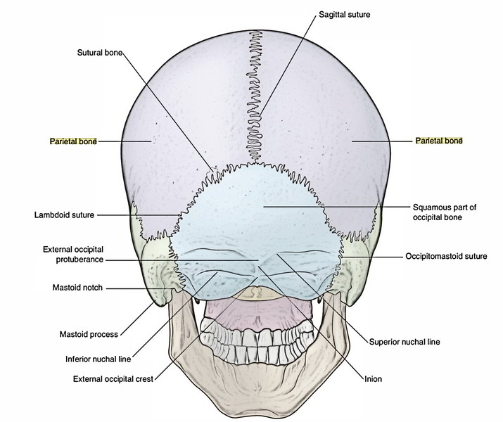
- Superior (sagittal) border: articulates with quite similar border of opposite parietal bone to create sagittal suture.
- Inferior (squamosal) border: articulates with 3 bones. From anterior to posterior, these are: (a) greater wing of sphenoid bone, (b) squamous part of temporal bone and (c) mastoid portion of the temporal bone.
- Anterior (frontal) border: articulates with all the frontal bone to create the coronal suture.
- Posterior (occipital) border: articulates with all the squamous part of the occipital bone to create the lambdoid suture.
Angles
The 4 angles of the parietal bone are as follows:
- Anteroinferior (sphenoidal) angle – is located at pterion
- Anterosuperior (frontal) angle – is located at the bregma
- Posteroinferior (mastoid) angle – is located at the asterion
- Posterosuperior (occipital) angle – is located at lambda
The anteroinferior (sphenoidal) angle projects down-wards and forwards to a substantial extent.
Clinical Significance
Sometimes, the parietal bone is split into 2 parts: upper and lower by an anomalous anteroposterior suture. The physician may mistake this state using a fracture. But it can be ruled out easily as the anomalous parietal suture is bilateral.
The regenerating ability of parietal bone is quite poor because of deficiency of cambium layer in the periosteum.

 (50 votes, average: 4.80 out of 5)