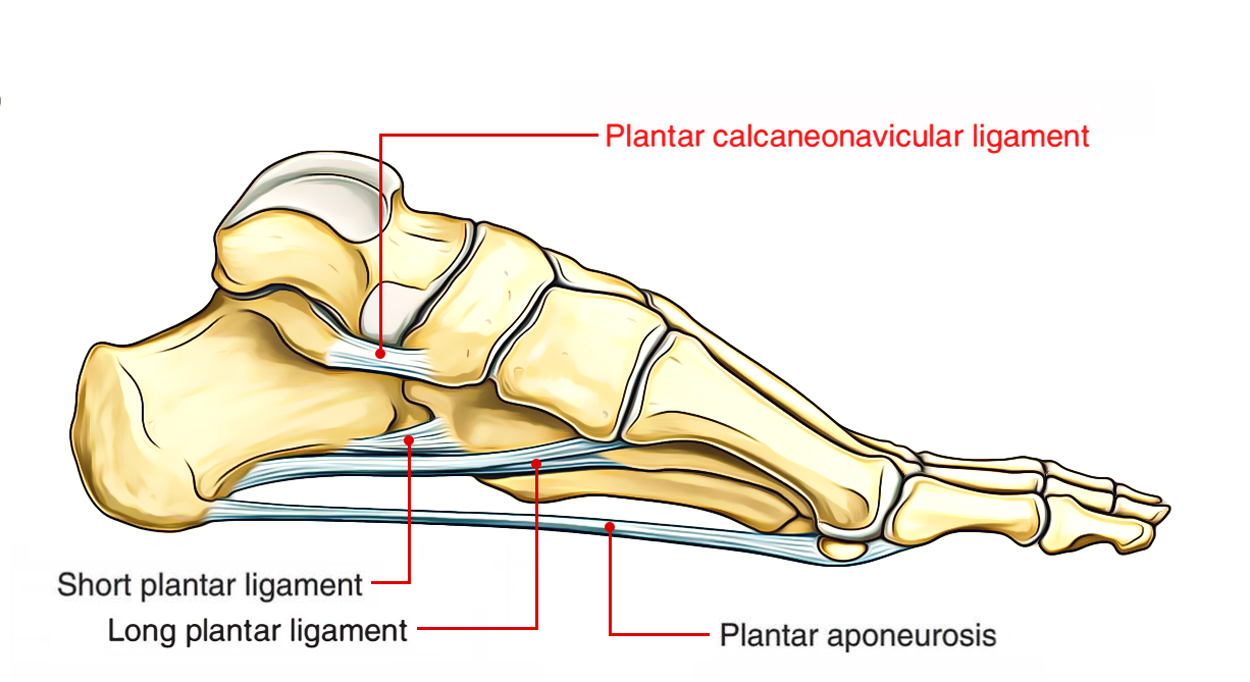
The spring ligament (plantar calcaneonavicular ligament) is a strong fibrocartilaginous band, which continues from the anterior margin of the sustentaculum tali to the plantar surface of navicular bone within its tuberosity and articular margin.
The plantar calcaneonavicular (spring) ligament complex is a group of ligaments that connect the calcaneum and navicular:
- Superomedial ligament
- Medioplantar oblique ligament
- Inferoplantar longitudinal ligament
The inferoplantar ligament is usually known as the lateral calcaneonavicular, and the medioplantar ligament is also known as the intermedial calcaneonavicular ligament.

Plantar Calcaneonavicular Ligament
It takes part in creating the socket for the head of the talus. It is the most essential ligament to maintain the medial longitudinal arch. Its superior surface has a triangular fibrocartilagenous aspect for the head of the talus.
Its plantar surface is reinforced by the tendon of tibialis posterior medially, and by tendons of flexor hallucis longus and flexor digitorum longus laterally. The talocalcaneonavicular joint allows the movements of inversion and eversion.
Surfaces
- This ligament not only serves to link the calcaneus and navicular, however also supports the head of the talus, forming part of the articular cavity in which it is received.
- The dorsal surface of the ligament presents a fibrocartilaginous facet, lined by synovial membrane, and upon this a part of the head of the talus rests.
- Its plantar surface is supported by the tendon of the Tibialis posterior
- Its median border is blended with the forepart of the deltoid ligament of the ankle-joint.
Structure
- The plantar calcaneonavicular ligament is a broad and thick strap of fibers, which links the anterior margin of the sustentaculum tali of the calcaneus to the plantar surface of the navicular.
- This ligament not just serves to connect the calcaneus and navicular, but supports the head of the talus, forming part of the articular cavity in which it is gotten.
- The dorsal surface of the ligament shows a fibrocartilaginous facet, outlined by the synovial membrane, and upon this a part of the head of the talus rests.
- Its plantar surface is strengthened by tendon of the tibialis posterior; its medial perimeter is combined with the forepart of the deltoid ligament of the ankle-joint.
- The plantar calcaneonavicular ligament helps to maintain the medial longitudinal arch of the foot, and by giving assistance to the head of the talus, bears the significant part of the body weight.
Clinical Significance
Flatfoot Deformity
This ligament plays an essential function in the advancement of acquired ‘flat foot defect’ (where the arch is missing) in adults. It has been linked to the stabilization of the foot’s longitudinal arch; this causes insufficiency of the spring ligament, which causes it to tear.
The plantar calcaneonavicular ligament complex is different in different feet. Sometimes it is comprised of two ligaments, the inferior calcaneonavicular and the superomedial; nevertheless, it usually has a third ligament.
Injury and Tear
Spring ligament injuries have a high connection with posterior tibial tendon tears. Spring ligament tears, just like posterior tibial tendon tears, are most generally seen in middle-aged women and are usually the result of long term deterioration. Disruption of the spring ligament destabilizes the longitudinal arch, allowing plantar and medial rotation of the head of the talus and valgus liganment of the calcaneus (pes planovalgus). The clinical outcome is an acquired flatfoot deformity.
Medical symptoms resemble those of posterior tibial dysfunction. Early in the illness procedure the patient might complain of unclear, activity-related discomfort at the medial ankle and foot or trouble with balance and strolling on uneven ground. Later, as the pes planovalgus defect advances, the patient frequently experiences activity related discomfort in the sinus tarsi and lateral malleolus, presumably due to impingement of lateral structures. With the advancement of subtalar and transverse tarsal osteoarthritis, the patient presents with pain, stiffness, and swelling.
Acute injuries of the spring ligament are scarce. Isolated tears of the spring ligament without associated PTT tear are extremely rare and can present as an acquired flatfoot deformity.

 (64 votes, average: 4.75 out of 5)