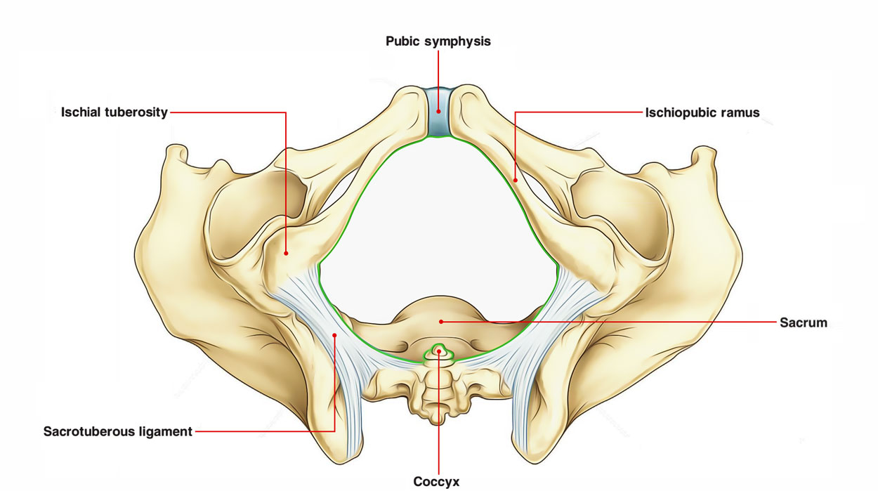
Every one of the joint’s surfaces is enveloped by hyaline cartilage and is connected throughout the midline to surrounding surfaces by fibrocartilage. The pubic symphysis is located anteriorly in between the surrounding surfaces of the pubic bones.
Structure of Pubic Symphysis
Functionally, the pubic symphysis is referred to as a secondary cartilaginous (fibrocartilage) joint and biomechanically, while starting as a synchondrosis, pubic symphysis becomes a nonsynovial amphi-arthrodial joint. The pubic symphysis joint includes two pubic bones (each including a pubic body and superior and inferior rami) and a stepping in fibrocartilaginous disc. The joint is bordered by interwoven layers of collagen fibers and the two significant ligaments related to it are:
- The superior pubic ligament, situated above the joint.
- The inferior pubic ligament, situated underneath it.

Pubic Symphysis
Development of Pubic Symphysis
- The centers of chondrification emerge in the primal scleroblastema and grow with each other in the midline, therefore creating the precursor of the pubic symphysis.
- A total cartilaginous pelvis exists by the second month of development.
- The symphysis then emerges at a point where the thick mesenchyme cavitates and separates in to hyaline cartilage and the fibrocartilaginous disc.
- Secondary centers of ossification take place at adolescence on the superior and inferior elements of the ventral surface of each pubic bone near the symphyseal face.
- Although irregular in development, have the tendency to unite and merge with the ventral border of each pubic bone in the early 20’s.
- The joint is wedge shaped, broader anteriorly than posteriorly, and the articular surfaces are oval.
- Pubic bodies are enveloped with hyaline cartilage. This cartilage is at first rather thick and starts to become thinner in the teenage years up until lastly just a small section is left in the adult (1 to 2mm).
Borders of Pubic Symphysis
- The osteocartilaginous border is at first smooth, and after that establishes irregularities (interdigitations) that gradually level after 23 to 25 years of age.
- A median cleft establishes in the postero-superior portion of the symphysial cleft in the second year of life.
- Secondary clefts that travelled along an eccentric course in between the hyaline cartilage covering the pubic bone and the fibrous cartilage, creating the symphysial disc and believed that those were traumatically customized clefts.
- When available, unlike primary clefts, these secondary clefts obtained a synovial lining.
- The symphysis has actually been referred to as being highly innervated with divisions via the pudendal and genitofemoral nerves.
- Its blood supply is stemmed from divisions of all significant vessels in the area consisting of the obturator, the internal pudendal, inferior epigastric, and medial circumflex arteries.
Ligaments of Pubic Symphysis
- The joint is held together by four ligaments (superior, inferior, anterior, and posterior) which create a constant circumferential envelope.
- The anterior ligament mixes with the rectus abdominis and adductor aponeurosis, and is the thickest and its decussating fibers involve in the pubic disc.
- The inferior (arcuate) ligament supports the most to the stability of the joint.
- Clefts and cysts exist within the ligaments.
Test Your Knowledge
PUBIC SYMPHYSIS

 (50 votes, average: 4.83 out of 5)