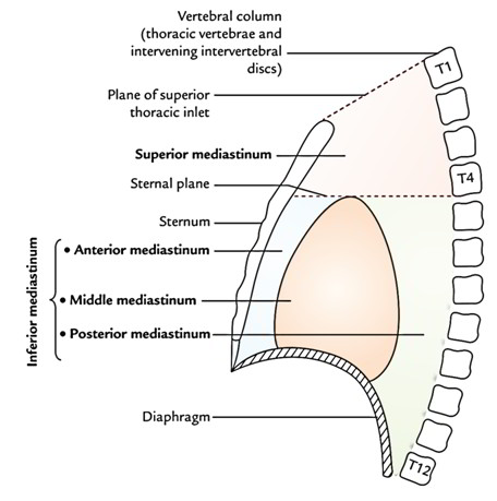
Boundaries
- Anterior: Manubrium sterni.
- Posterior: Bodies of upper 4 thoracic vertebrae.
- Superior: Plane of superior thoracic aperture.
- Inferior: An imaginary plane going through the sternal angle in front and lower border of the body of fourth thoracic vertebra behind (transverse thoracic plane).
- On every side: Mediastinal pleura.
Contents
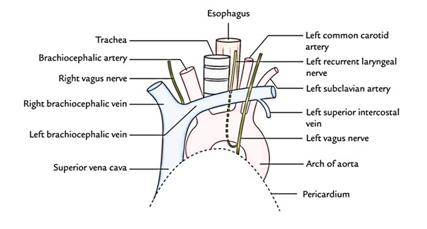
Arteries
(a) Arch of aorta.
(b) Brachiocephalic artery.
(c) Left common carotid artery.
(d) Left subclavian artery.
Veins
(a) left and right brachiocephalic veins.
(b) Upper half of the superior vena cava (SVC).
(c) Left superior intercostal vein.
Nerves
(a) Phrenic nerves (left and right).
(b) Vagus nerves (left and right).
(c) Sympathetic trunks and cardiac nerves (left and right).
(d) Left recurrent laryngeal nerves.
Lymphoid Organs And Lymphatics
(a) Lymph nodes.
(b) Thoracic duct.
(c) Thymus.
Tubes
(a) Trachea.
(b) Esophagus.
Muscles
(a) Sternohyoid.
(b) Sternothyroid.
(c) Longuscolli.
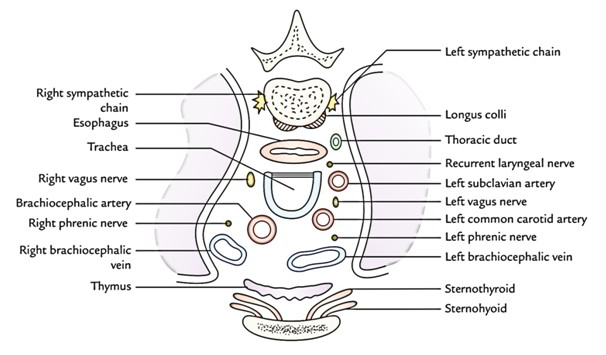
For the purpose of orientation during surgery, the major structures of superior mediastinum are arranged in the following order from anterior to posterior:
1. Thymus.
2. Large veins.
3. Large arteries.
4. Trachea.
5. Esophagus and thoracic duct.
6. Sympathetic trunks.
Clinical Significance
Potential dead space in superior mediastinum: In superior mediastinum, all large veins (superior vena cava, left and right brachiocephalic veins) are on the right side and all the large arteries (arch of aorta and its 3 branches) are on the left side. Consequently, during increased blood flow, the large veins expand enormously while the large arteries don’t expand at all. This is because there’s sufficient dead space on the right side. It’s into this space that tumors of mediastinum tend to project.

 (49 votes, average: 4.65 out of 5)