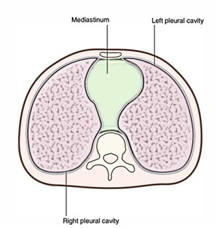
The Mediastinum is a compartment located between two pleural sacs. It goes from the sternum anteriorly to the thoracic vertebrae posteriorly. Also it goes from the superior thoracic aperture to the inferior thoracic aperture. It is a thick midline partition.
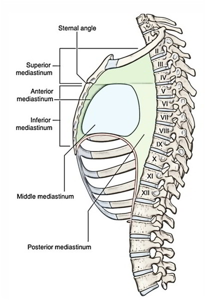
A horizontal plane going through the sternal angle and the intervertebral disc between vertebrae TIV and TV splits the mediastinum into superior and inferior parts. The inferior part is further subdivided by the pericardium, which encloses the pericardial cavity enclosing the heart. The pericardium and heart make up the middle mediastinum.
The anterior mediastinum is located between the sternum and the pericardium; the posterior mediastinum is located between the pericardium and thoracic vertebrae.
Boundaries
- Anterior: Sternum.
- Posterior: Vertebral column (bodies of thoracic vertebraeand intervening intervertebral discs).
- Superior: Superior thoracic aperture.
- Inferior: Diaphragm.
- On every side: Mediastinal pleura.
The mediastinum isn’t a rigid structure as observed by the students in the cadaver (embalmed dead body). In a living individual, mediastinum is a highly mobile septum for the reason that it consists primarily of hollow visceral structures bound together by loose connective tissue, frequently infiltrated with fat.
Contents
The major contents of mediastinum are:
- Thymus.
- Heart enclosed in the pericardial sac.
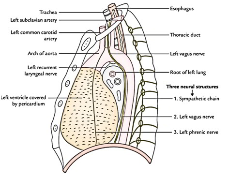
- Major arteries and veins like thoracic aorta, pulmonary trunk, etc.
- Trachea.
- Esophagus.
- Thoracic duct.
- Neural structures, like sympathetic trunks, vagus nerves, phrenic nerve, etc.
- Lymph nodes.
Divisions
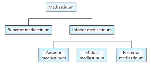
For the purpose of description and organization of structures the mediastinum is artificially split into 2 parts:
1. Superior mediastinum and inferior mediastinum by an imaginary plane (transverse thoracic plane) going through the sternal angle anteriorly, and lower border of the body of the fourth thoracic (T4) vertebra/intervertebral disc T4 and T5 vertebrae posteriorly.
2. The inferior mediastinum is further subdivided into 3 parts by the pericardium (enclosing heart). The part in front of the pericardium is referred to as anterior mediastinum, and the part supporting the pericardium is referred to as posterior mediastinum. The pericardium and its contents (heart and roots of its great vessels) constitute the middle mediastinum.
Clinical Significance
Visualization Of Subdivisions Of Mediastinum In Chest Radiograph
The subdivisions of mediastinum are well-appreciated in lateral view of X-ray chest. The shadow above the sternal plane represents superior mediastinum.
The subdivisions of inferior mediastinum are demarcated as under:
- 1. Cardiac shadow above the anterior part of diaphragm represents the middle mediastinum.
- 2. Retrosternal space/space in the front of cardiac shadow represents the anterior mediastinum.
- 3. Retrocardiac space/space between the cardiac shadow and shadow of vertebral column represents the posterior mediastinum.
Mediastinitis
It’s the inflammation of the loose connective tissue of the mediastinum. The fascial spaces of the neck (example: pretracheal and retropharyngeal spaces) extend below into the mediastinum inside the thoracic cavity. For that reason, deep infections of the neck may readily spread into the thoracic cavity causing mediastinitis.
Subcutaneous Emphysema In The Root Of Neck
The structures that make up mediastinum are embedded in the loose connective tissue that is continuous with the loose connective tissue of the root of the neck. For that reason, in esophageal perforations caused by penetrating wounds, air escapes into the connective tissue spaces of mediastinum and could ascend below the fascia to the root of the neck producing subcutaneous emphysema.
Mediastinal Syndrome
The compression of mediastinal structures by any growth like tumor or cyst gives rise to a group of signs and symptoms, producing a clinical condition referred to as mediastinal syndrome.
The common causes of mediastinal syndrome are bronchiogenic carcinoma, aneurysm of aorta, enlargement of mediastinal lymph nodes in Hodgkin disease.
The clinical features of mediastinal syndrome are:
- engorgement of veins in the upper half of the body: because of obstruction of SVC,
- dyspnea (difficulty in breathing): because of compression of trachea,
- dysphagia (difficulty in swallowing): because of compression of esophagus,
- dyspnea (hoarseness of voice): because of compression of left recurrent laryngeal nerve, and
- erosion of bodies of thoracic vertebrae: because of pressure on the vertebral column
Widening Of The Mediastinum
The widening of mediastinum is frequently observed in chest radiographs. It can happen because of a number of reasons, like (a) hemorrhage into the mediastinum from lacerated great vessels (aorta, SVC) following trauma, example: head on collision, massive enlargement of mediastinal lymph nodes because of cancer of lymphoid tissue, viz. malignant lymphoma, and enlargement (hypertrophy) of heart because of congestive heart failure (CHF). It’s the common cause of widening of inferior mediastinum.
Mediastinal Shift
The mediastinal shift is common in lung and pleural pathology. The mediastinum shifts to the affected (diseased) side because of appreciable reduction in lung volume and decrease in intrapleural pressure- as in collapse of lung and atelectasis. The mediastinum shifts to the healthy side when the intrapleural pressure is appreciably high on the affected side – as in pneumothorax and hydrothorax.
Mediastinal shift indicates lung pathology. The mediastinal shift can be discovered by palpating the trachea in the suprasternal notch.
Extension Of Pus Into The Posterior Mediastinum From Neck
The posterior mediastinum is continuous via superior mediastinum with spaces in the neck between pretracheal and prevertebral layers of deep cervical fascia like retropharyngeal space, etc. For That Reason, pus from neck can extend into the posterior mediastinum.
Extension Of Pus Into The Thighs From Posterior Mediastinum.
The psoas sheath interacts together with the posterior mediastinum by a funnel-shaped orifice. For that reason, pus from posterior mediastinum can easily goes into the psoas sheath and tracks down in the thighs in the region of femoral triangle.

 (52 votes, average: 4.87 out of 5)