The Ankle Joint is a powerful weight bearing joint of the lower limb. The ankle supports the weight of entire body and also remains mostly active in day-to-day life in a lot of physical activities. The ankle also helps in maintaining body posture.
Ankle Joint
Type
The ankle joint or talocrural joint is a synovial joint of hinge type and is the only mortise and tenon joint in the human body.
Articular Surfaces
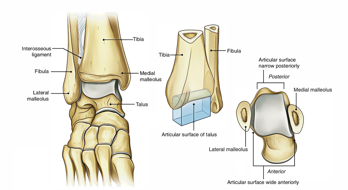
Ankle Joint: Articular Surfaces
The proximal articular outermost layer of the ankle joint is made by the articular facets of the:
- The lower end of tibia consisting of its medial malleolus.
- Lateral malleolus.
- Inferior transverse tibiofibular ligament.
These three together create a deep tibiofibular socket which is also termed as tibiofibular mortise.
The distal articular outermost layer of the talocrural joint is composed of articular facets on the upper, medial, and lateral aspects of the body of the talus.
The body of the talus bone presents three articular surfaces:
- Superior pulley-shaped articular surface (trochlear surface).
- Medial comma-shaped articular surface.
- Lateral triangular articular surface.
The wedge-shaped body of the talus fits into the tibiofibular socket.
Relations
The articular surface on the inferior aspect of the lower end of tibia articulates with trochlear surface of the talus.
The articular surface on the lateral aspect of medial malleolus articulates with the articular surface on the medial side of the talus.
The articular surface on the medial aspect of lateral malleolus articulates with all the large triangular articular surface on the lateral side of the body of talus.
The ankle joint resembles a pincer or monkey wrench grasping a portion of hemisphere. (cf. tibiofibular mortise grasping the wedge shaped body of the talus.).
The Stability of the Ankle Joint
The trochlear surface on the superior aspect of the body of talus is wider in front than behind.
During dorsiflexion, ankle joint of the anterior wider part of the trochlea moves posteriorly and fits correctly into the tibiofibular mortise (pincer), thus joint is stable. During plantar flexion, the narrow posterior part of the trochlea doesn’t fit correctly in the tibiofibular mortise (pincer), thus the joint is unstable during plantar flexion.
Variables Keeping the Solidity of the Ankle Joint
- The close interlocking of its articular surfaces.
- Strong, medial, and lateral collateral ligaments.
- Deepening of tibiofibular socket posteriorly by the inferior transverse tibiofibular ligament.
- Tendons (4 in front and 5 behind) crossing the ankle joint.
- Other ligaments of the joint.
Ligaments
The essential ligaments of the ankle joint are:
- Capsular ligament.
- Medial and lateral collateral ligaments.
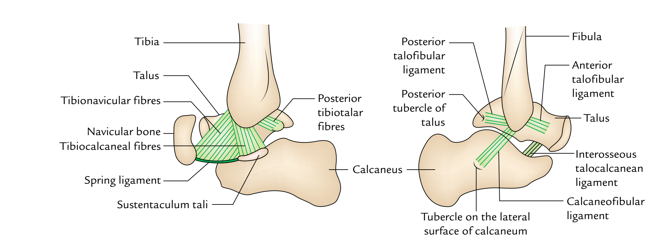
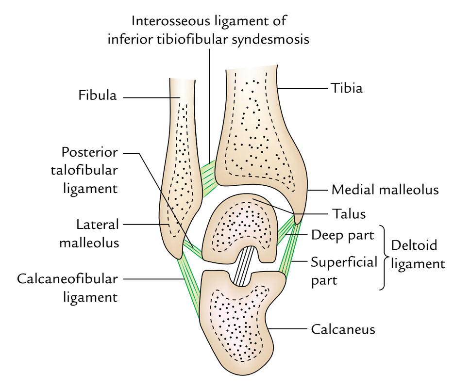
Ankle Joint: Ligaments
Fibrous Capsule
It encompasses the joint entirely. It’s connected to the articular margins of the joint all around with 2 exceptions:
- Posterosuperiorly it’s connected to the inferior transverse tibiofibular ligament.
- Anteroinferiorly it’s connected to the dorsum of the neck of talus at some distance from the trochlear surface.
The joint capsule is thin in front and behind to enable hinge movements and thick on either side where it combines with the collateral ligaments.
The synovial membrane lines the inner surface of the joint capsule, but ends at the periphery of the articular cartilages. A small synovial process goes upward into the inferior tibiofibular syndesmosis.
Deltoid or Medial Ligament
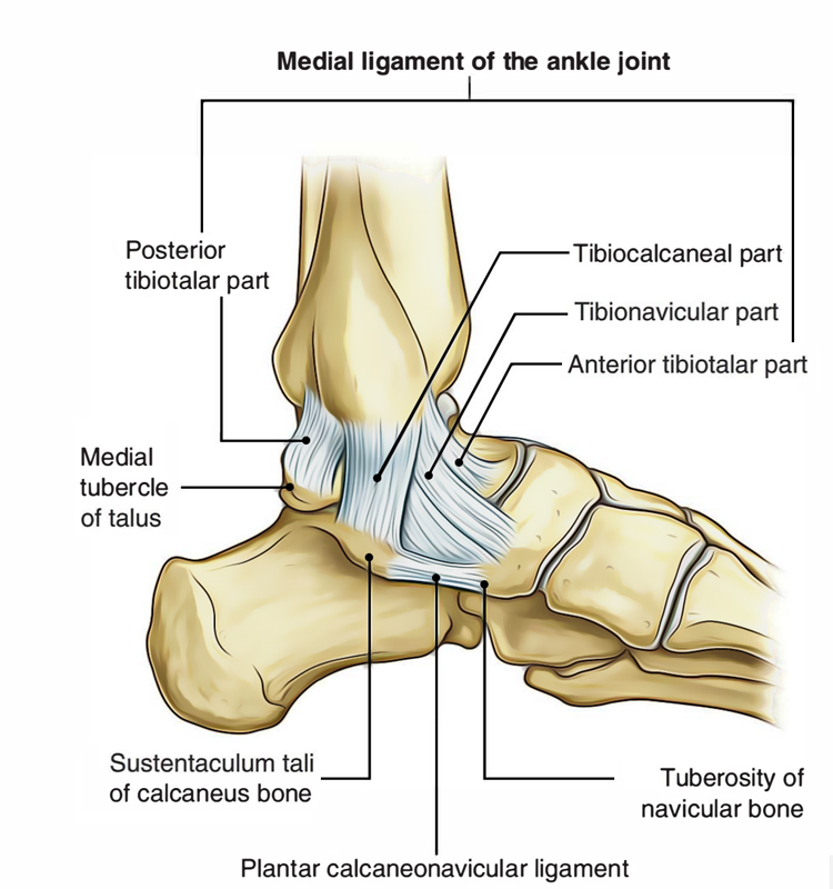
Ankle Joint: Medial Ligament
The deltoid ligament is an extremely strong and triangular shaped ligament on the medial side of the ankle. It’s split into two parts: superficial and deep.
Above, both the parts have a common connection to the apex and margins of the medial malleolus. Below, the connection of superficial and deep parts differs as under:
The superficial parts fibres are split into three parts: anterior, middle, and posterior.
- Anterior fibres (tibionavicular) are connected to the tuberosity of navicular bone and the medial margin of spring ligament.
- Middle fibres (tibiocalcanean) are connected to the entire length of sustentaculum tali.
- Posterior fibres (posterior tibiotalar) to the medial tubercle and adjoining part of the medial surface of talus.
The Deep part (anterior tibiotalar) is connected to the anterior part of the medial surface of talus.
Lateral Ligament
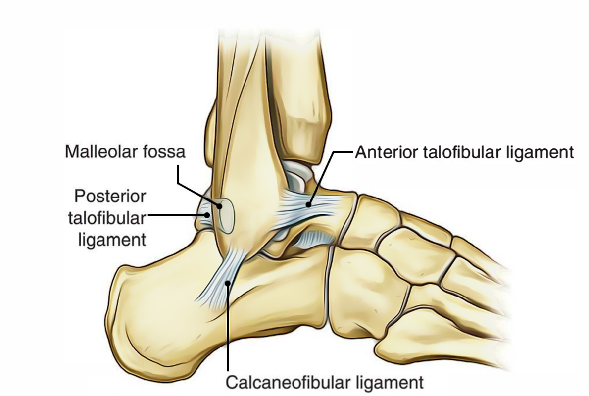
Ankle Joint: Lateral Ligament
The lateral ligament is composed of three parts: anterior talofibular, posterior talofibular, and calcaneofibular.
- Anterior talofibular ligament is a poor flat band, which goes forwards and medially from the anterior margin of the lateral malleolus, to the neck of talus just in front of the fibular facet.
- Posterior talofibular ligament is a powerful band, which goes backwards and medially from the posterior margin of the lateral malleolus to the posterior tubercle of the talus.
- Calcaneofibular ligament is a long rounded cord, which runs downward and backward from the pass on the lower border of lateral malleolus to the tubercle on the lateral surface of the calcaneum.
Superficially, the deltoid ligament is crossed by the tendons of tibialis posterior and flexor digitorum longus on the other hand lateral ligament is crossed superficially by the tendons of peroneus longus and brevis.
Relations of the Ankle Joint
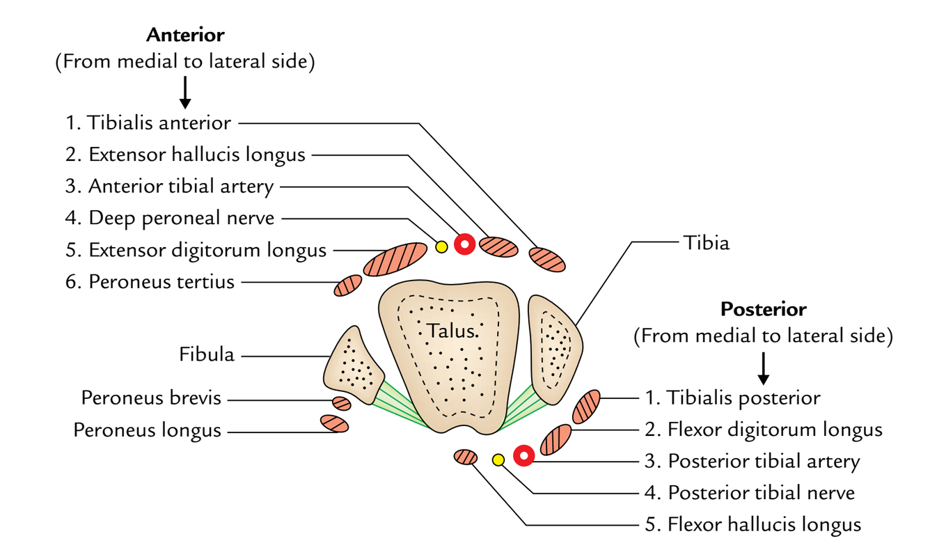
Ankle Joint: Relations
Anterior: Anteriorly from medial to lateral side the ankle joint is related to these structures:
- Tibialis anterior.
- Extensor Hallucis longus.
- Anterior tibial Artery.
- Deep peroneal Nerve.
- Extensor Digitorum longus.
- Peroneus tertius.
Mnemonic: The Himalayas Are Not Dry Tablelands.
Posterior: Posteriorly from medial to lateral side the ankle joint is related to these structures:
- Tibialis posterior.
- Flexion Digitorum longus.
- Posterio r tib ial Artery.
- Posterio r tib ial N erve.
- Flexor Hallucis longus.
Mnemonic: The Doctors Are Not Here.
Arterial Supply
The ankle joint is supplied by the malleolar branches of anterior tibial, posterior tibial, and peroneal arteries.
Nerve Supply
The ankle joint is innervated by the branches of deep peroneal and tibial nerves. (The segmental innervations is by L4, L5; S1, S2 spinal sections).
Movements
The following movements take place in the ankle joint:
- Dorsiflexion.
- Plantar flexion.
When the foot is plantar flexed, the ankle joint also allows some movements of side to side gliding, rotation, adduction, and abduction.
Dorsiflexion
In dorsiflexion, the forefoot is lifted and the angle between the front of the leg and dorsum of the foot is decreased. In this movement, the wider anterior part of trochlea is compelled posteriorly between the malleoli. It’s a close packed position of the ankle joint with maximum congruence of the articular surfaces and tension of ligaments. The ankle joint is the most stable in dorsiflexion.
Plantar Flexion
- In plantar flexion, the forefoot is depressed, and the angle between the leg and foot is raised. In this position, the tibiofibular socket encloses the narrower posterior part of the trochlear surface of the talus and some joint space isavailable between the tibiofibular mortise and the narrow posterior part of the trochlea. It’s a loose filled position of the ankle joint. The joint is shaky in plantar flexion.
- The ankle joint is stable in dorsiflexion and shaky in plantar flexion.
- The range of motion (ROM) for dorsiflexion is 20 ° and for plantar flexion is 45 °.
- The dorsiflexion is restrained by the L4, L5 spinal segments and plantar flexion by the S1, S2 spinal sections.
Movements of the Ankle Joint
| Muscles producing movements | ||
|---|---|---|
| Movements | Principal muscles | Accessory muscles |
| Dorsiflexion | Tibialis anterior | 1.Extensor digitorum longus. |
| 2.Extensor hallucis longus. | ||
| 3.Peroneus tertius. | ||
| Plantar flexion | 1.Gastrocnemius. | 1.Plantaris. |
| 2.Soleus. | 2.Tibialis posterior. | |
| 3.Flexor hallucis longus. | ||
| 4.Flexor digitorum longus. |
Clinical Significance
Ankle Sprains
- The excessive stretches and/or tearing of ligaments of the ankle joint is named the ankle sprain. The ankle sprains are normally caused by the falls from height or spins of ankle.
- When the plantar-flexed foot is excessively inverted, the anterior and posterior talofibular and calcaneofibular ligaments are stretched and torn. The anterior talofibular ligament is most commonly torn.
- When the plantar-flexed foot is excessively everted, the deltoid ligament isn’t torn; instead there’s an avulsion fracture of medial malleolus.
- The inversion sprains are much more common than eversion sprains.
Dislocation of the Ankle
The dislocations of ankle joint are uncommon because it’s a quite stable joint because of tibiofibular mortise. Nevertheless, whenever dislocation takes place it’s constantly escorted by the fracture of 1 of the malleoli.
Pott’s Fracture (Fracture Dislocation of the Ankle)
It takes place when the foot is caught in the rabbit hole and everted forcibly. In this state, the subsequent series of events happens:
- Oblique fracture of the lateral malleolus because of internal rotation of the tibia.
- Transverse fracture of the medial malleolus as a result of pull by powerful deltoid ligament.
- Fracture of the posterior margin of the lower end of tibia (third malleolus) because it’s carried forward. These phases are also referred to as first, 2nd, and third degree of Pott’s fracture, respectively. The third degree of Potts fracture is also termed trimalleolar fracture.
Optimum Position of the Ankle
The optimum position of the ankle is one where ankle joint is in slight plantar flexion. The understanding of position is important for using plaster cast in the ankle region.
Test Your Knowledge
Ankle Joint

 (58 votes, average: 4.14 out of 5)