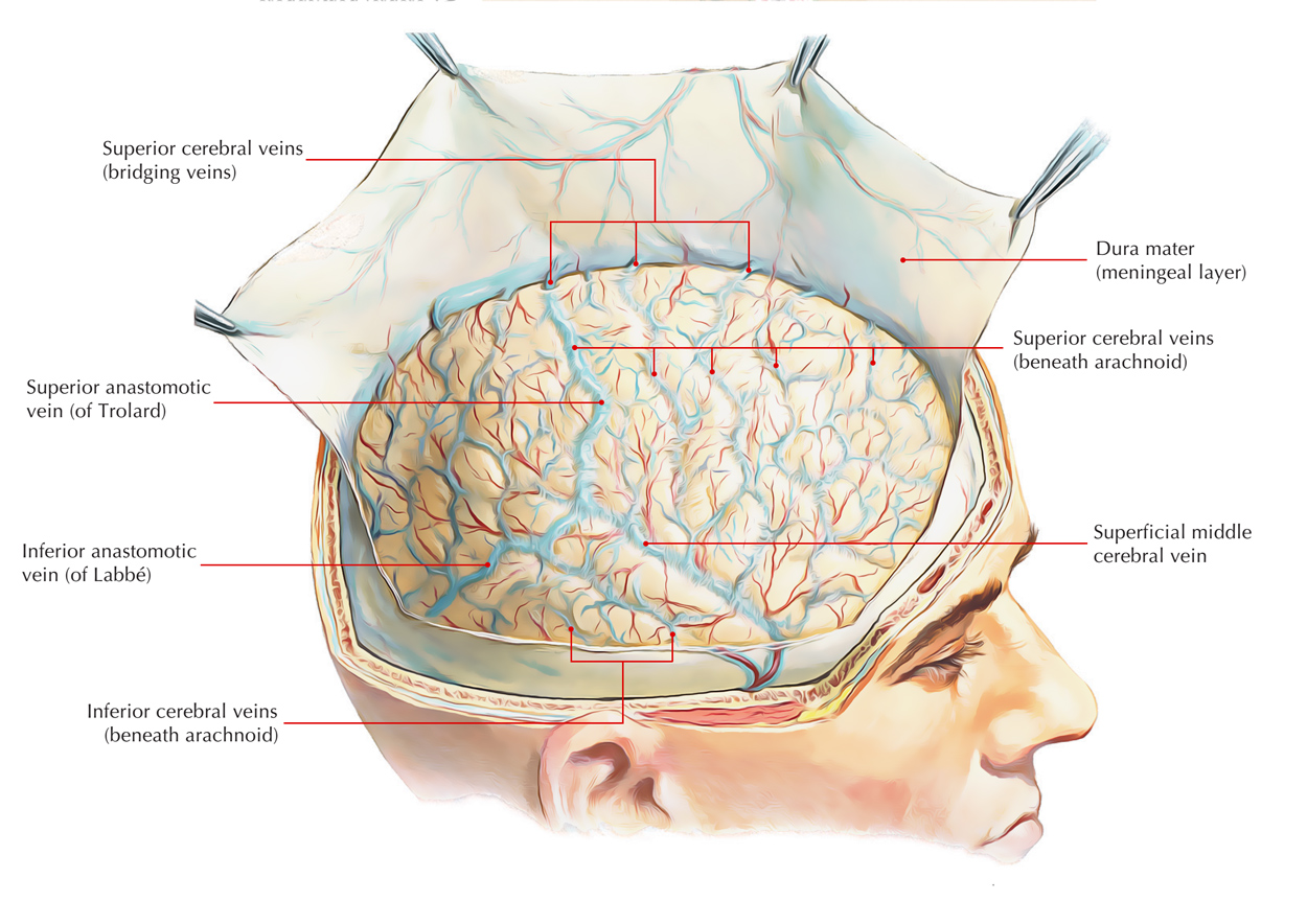
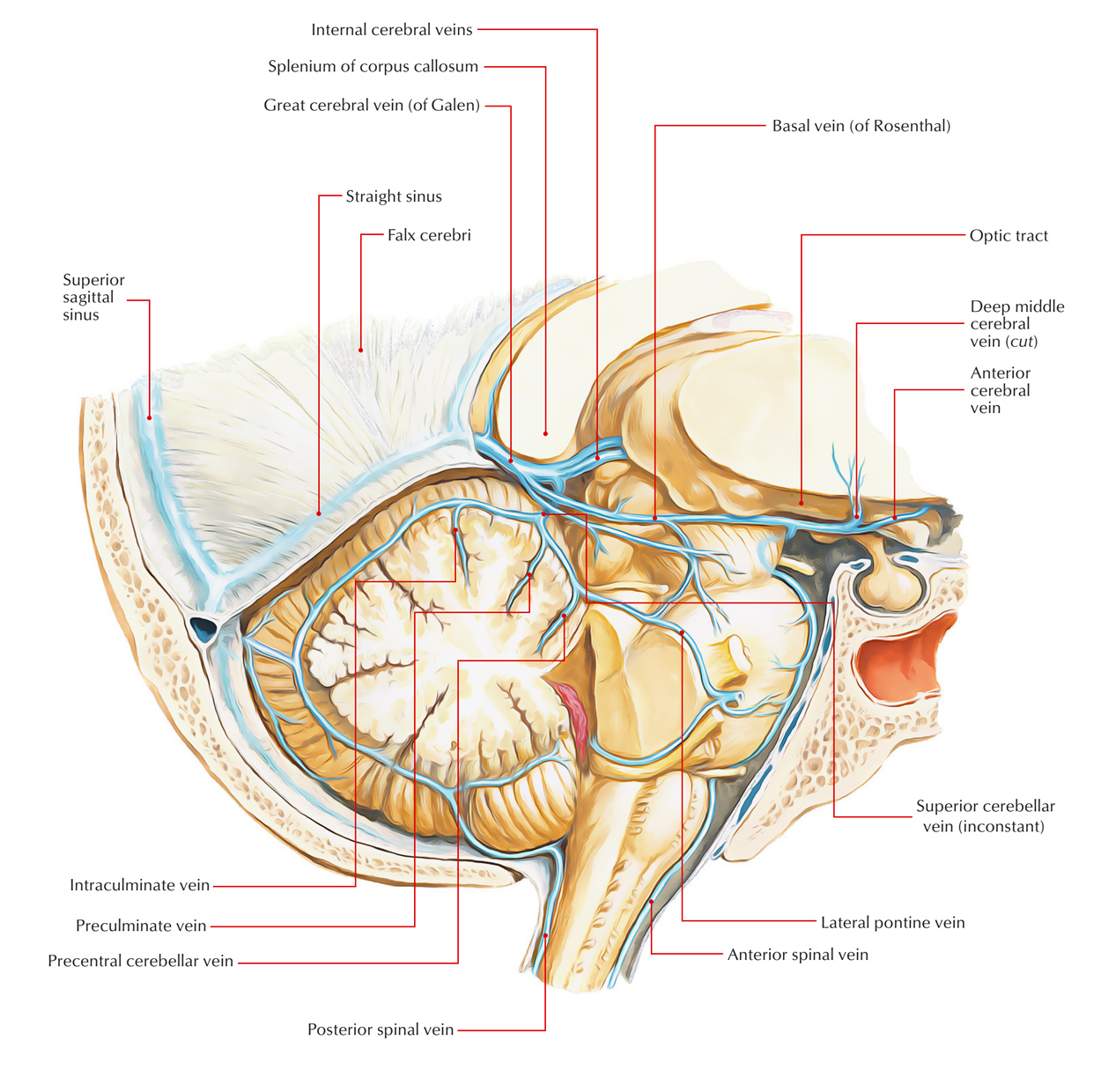
The cerebral veins are found within the subarachnoid space and withdraw deoxygenated blood from the brain parenchyma. The cerebral veins drain the external surfaces and the internal zones of the cerebral hemisphere. They perforate the meninges as well as channel further within the cranial venous sinuses. They are categorized within external (superficial) and internal cerebral veins.
External Cerebral Veins
The surface/cortex of the hemisphere is emptied by the external cerebral veins. They are divided in three groups:
- Superior
- Middle
- Inferior

External Cerebral Veins
Superior Cerebral Veins
Superior cerebral veins drain the upper portions of the superolateral as well as medial surfaces of the cerebral hemisphere. They rise upwards, penetrate the arachnoid mater, and afterwards, in order to enter the superior sagittal sinus cross the subdural space.
The anterior veins open at right angle, whereas the posterior open obliquely opposite towards the flow of bloodstream in the superior sagittal sinus, thus inhibiting their collapse by growing CSF pressure.
Middle Cerebral Veins
There are four middle cerebral veins; two are present on each side: superficial middle cerebral vein and deep middle cerebral vein.
The superficial middle cerebral vein is superficially located within the lateral sulcus.
Posteriorly, it connects with the superior sagittal sinus through superior anastomotic vein (of Trolard) along with the transverse sinus by inferior anastomotic vein (of Labbe), whereas anteriorly, it travels forwards in order to drain inside the cavernous sinus.
The deep middle cerebral vein is located deep within the lateral sulcus on the insula along with middle cerebral artery. It travels downwards as well as forwards and for creating the basal vein, it connects with the anterior cerebral vein.
Inferior cerebral veins
The inferior cerebral veins are smaller in size but numerous. They empty the inferior surface and lower parts of medial and superolateral surfaces of the cerebral hemisphere within neighboring intracranial dural venous sinuses.
Other Veins
Anterior Cerebral Vein
Nearby the corpus callosum, it goes along with the anterior cerebral artery and the parts of medial surface emptied by it, which cannot be drained into the superior and inferior sagittal sinuses.
Basal Vein (of Rosenthal)
It is created by the combination of three veins: anterior cerebral, deep middle cerebral and striate veins, in the section of anterior perforated substance at the base of the brain. The striate veins arise from the anterior perforated substance.
The basal vein travels posteriorly around the midbrain, medial towards the uncus and parahippocampus, and ends under the splenium of corpus callosum within the great cerebral vein (of Galen).
Apart from the three forming veins, the basal vein obtains the branches from:
- Cerebral peduncle.
- Uncus and parahippocampus.
- Structures of interpeduncular fossa.
- Optic tract and olfactory trigone.
- Inferior horn of the lateral ventricle.
Internal Cerebral Veins
Through the combination of three veins: thalamostriate, septal, and choroidal, all internal cerebral veins are created on the Interventricular foramen of Monro.

Internal Cerebral Vein
The two internal cerebral veins in the middle of the two layers of tela choroidea of 3rd ventricle, travel posteriorly. One through each side of midline and afterwards unite together beneath the splenium of corpus callosum in order to create the great cerebral vein (of Galen), which empties within the straight sinus.
Among the deep veins of the cerebrum, the most essential are thalamostriate, septal, and choroidal veins:
- The Thalamostriate a.k.a. striothalamic vein empties the thalamus along with basal ganglia
- The Septal vein empties the septum pellucidum
- The Choroidal vein drains the choroids plexus
Great Cerebral Vein (of Galen)
Great cerebral vein is a single vein of approximately 2 cm lengthwise. Two internal cerebral veins combine below and behind the splenium of corpus callosum in order to create great cerebral vein. It instantaneously gets the two basal veins and it connects with the inferior sagittal sinus in order to create the straight sinus after a short backward course.
The constituent veins of the great cerebral vein are:
- Internal cerebral veins.
- Basal veins.
- Veins from colliculi, i.e., tectum of midbrain.
- Veins from cerebellum along with adjacent portions of the occipital lobes of the cerebrum.

 (56 votes, average: 4.64 out of 5)