These joints take place between occipital condyles, atlas and axis.
All these joints are superbly adapted for eye and head coordination and serve as a universal joint and together allow horizontal and vertical scanning movements of the head. The craniovertebral joints contain:
- Atlanto-occipital joints.
- Atlantoaxial joints.
Atlanto-Occipital Joints
These are 2 atlanto-occipital joints, 1 on either side between the atlas vertebra and occipital bone.
Type
All these are synovial joints of ellipsoidal variety.
Articular Surfaces
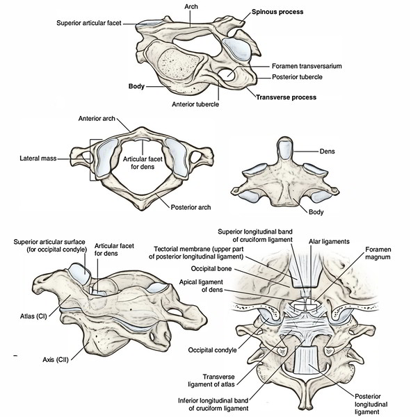
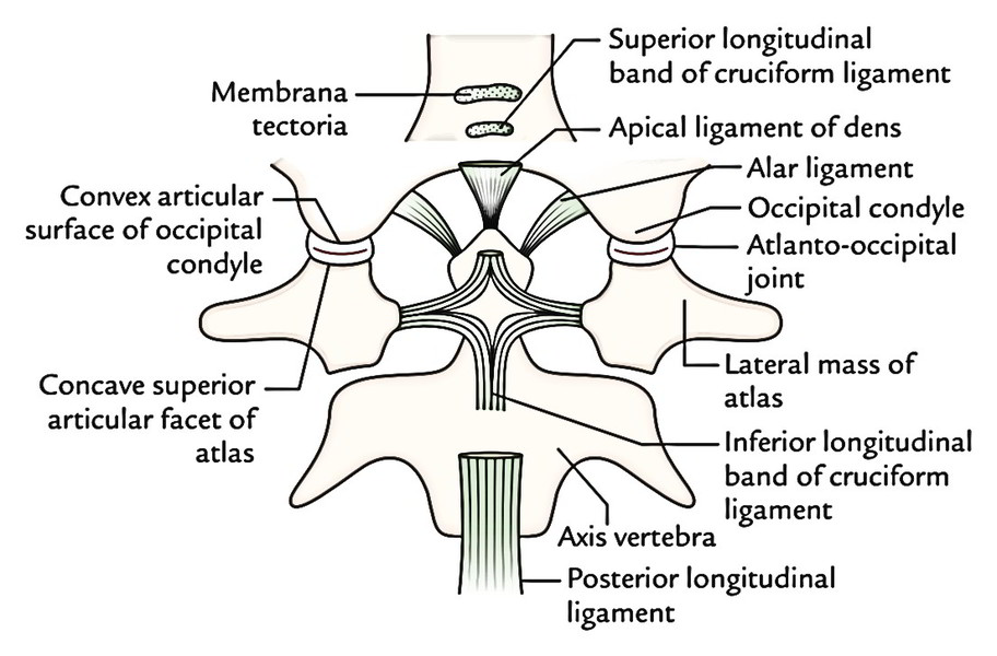
Above: Convex articular surface of occipital condyles.
Below: Concave superior articular facets of the atlas (cervical vertebra). With the constrictions in the middle, they can be elongated and kidney shaped. They may be directed medially and forwards. So the superior and inferior articular surfaces are reciprocally arch.
Ligaments
Fibrous capsule (capsular ligament): It encompasses the joint and is connected to the margins of the articular surfaces. It is thick posterolaterally and thin posteromedially.
Accessory ligaments
- Anterior atlanto-occipital membrane
- It’s connected below to the anterior arch of the atlas and above to the anterior margin of the foramen magnum.
- It fuses with the fibrous capsule laterally and is fortified by cord-like anterior longitudinal ligament anteriorly.
Posterior atlanto-occipital membrane
It’s connected below to the upper border of the posterior arch of the atlas and above to the posterior margin of the foramen magnum. Inferolaterally it arches over a groove on the upper surface of the posterior arch of atlas for vertebral artery and first cervical nerve.
Arterial Supply
By vertebral artery
Nerve Supply
By first cervical nerve.
Movements
Since these are ellipsoid joints, the movements are allowed in transverse as well as in anteroposterior axes.
Movements happening at atlanto-occipital joints.
| Movement | Axis | Muscles responsible for movements |
|---|---|---|
| Flexion and extension (nodding or yes movements) | Transverse | Flexion by: longus capitis and rectus capitis anterior Extension by: Rectus capitis posterior major and minor Semispinalis capitis Splenius capitis Upper part of the trapezius |
| Lateral flexion (slight) | Anteroposterior | Rectus capitis lateralis |
The line of gravity of the weight of the head (about 7 pounds) enters in front of the atlanto-occipital joints. For that reason, position of head in erect position is kept by the traction caused by the extensor muscles, especially by semispinalis capitis. Therefore semispinalis muscle accounts for keeping the head in proper position.
Atlantoaxial Joints
3 well dislocated joints are created between the atlas and the axis, viz.
- Median atlantoaxial joint.
- 2 lateral atlantoaxial joints.
All these joints function as 1 unit to generate the movement of rotation of atlas with head around the vertical axis.
Median Atlantoaxial Joint
Type
It’s a synovial joint of pivot variety. It’s created between the holes of axis and osseoligamentous ring created by anterior arch and transverse ligament of the atlas.
Articular Surfaces
Odontoid process of axis.
Anterior arch and transverse ligament of atlas.
- There are 2 joints- anterior and posterior- with individual synovial cavities.
Anteriorly, the vertically oriented oval facet on the anterior surface of lairs articulates with a quite similar facet on the posterior surface of the anterior arch of the atlas.
Posteriorly, the cartilaginous anterior surface of transverse ligament of atlas articulates with the horizontally oriented ovoid facet on the posterior outermost layer of the base of the holese.
Ligaments
Fibrous capsule: It’s loose and encompasses the anterior joint. It’s connected around the margins of the articular facets. It’s lined by synovial membrane.
Transverse ligament: It’s connected on every side to the medial surface of the lateral mass of the atlas. In the median plane its fibres are prolonged: (a) upwards to the basiocciput and (b) downwards to the body of the axis, hence creating the cruciform ligament of the atlas. The transverse ligament covers the narrow neck of the lairs and prevents its backward dislocation. A synovial bursa is interposed between the transverse ligament and lairs. It’s said to be the large posterior part of the median atlantoaxial joint.
Ligaments joining the axis together with the occipital bone
Apical ligament of the lairs goes from tip of odontoid process to the upper surface of the basilar part of the occipital bone near the foramen magnum.
Morphologically it signifies the remnant of the notochord (cf. Nucleus pulposus).
Alar ligaments, 1 on every side, stretch from the upper part of the lairs (on side of the tip) to the tubercle on the medial aspect of the occipital condyle. These ligaments are extremely formidable and check excessive rotation and flexion of head. They’re, consequently, termed check ligaments.
Membrana tectoria is an upward continuation of the posterior longitudinal ligament. It is located posterior to the transverse ligament of the atlas. Inferiorly, it’s connected to the posterior outermost layer of the body of the axis and superiorly to the upper surface of the basilar part of the occipital bone above the connection of upper band of the cruciform ligament.
Lateral Atlantoaxial Joints
These are matched, 1 on every side.
Type
Synovial joint of plane variety.
Articular Surfaces
Above: Inferior facet of the lateral mass of the atlas. It’s flat.
Below: Superior articular facet of axis. It’s also flat consequently superior and inferior articular facets are reciprocally arch.
Capsules: It’s connected to the margin of articular surfaces. It’s supported by anterior longitudinal ligament and ligamentum flavum.
Movements
All these are simultaneous at all the 3 atlantoaxial joints and consist just about completely of rotation of axis. In reality, the osseoligamentous ring of atlas supporting the atlas rotates around the central pivot created by the odontoid process. The rotation is restricted primarily by alar ligaments. Rotatory movements of atlas with head, around vertical axis are also referred to as No movements.
Clinical Significance
Hangman’s fracture: It’s defined by the fracture of the pedicles of the axis vertebra. It’s a critical extension injury of the neck that takes place from automobile accident or a fall from height. Since in this injury the vertebral canal is enlarged because of forward displacement of the body of axis, the spinal cord is seldom compressed.
This injury is thus termed because during execution by hanging, the knot of Hangman’s rope underneath the chin causes unexpected intense extension injury of the neck.
Executive hanging can also cause fracture of odontoid process or separation of axis from the third cervical vertebrae.
The fractures of odontoid process of axis vertebra arerelatively common and typically takes place because of fall or blow on the head. Excessive freedom of the fractured odontoid process, especially if related to rupture of transverse ligament of atlas can cause compression of the spinal cord.
In executing hanging, death happens because of posterior dislocation of odontoid process (following rupture of transverse ligament of atlas) smashing the lower part of medulla oblongata which places essential centers and adjoining part of the spinal cord.

 (57 votes, average: 4.54 out of 5)