The Cubital Fossa is corresponding or homologous to the popliteal fossa of the lower limb. It is a triangular hollow in front of the elbow.
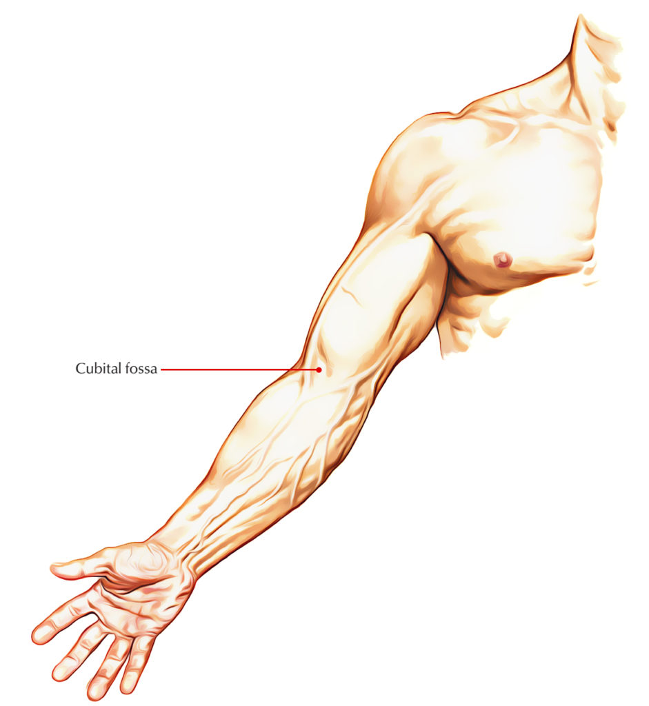
Cubital Fossa
Borders
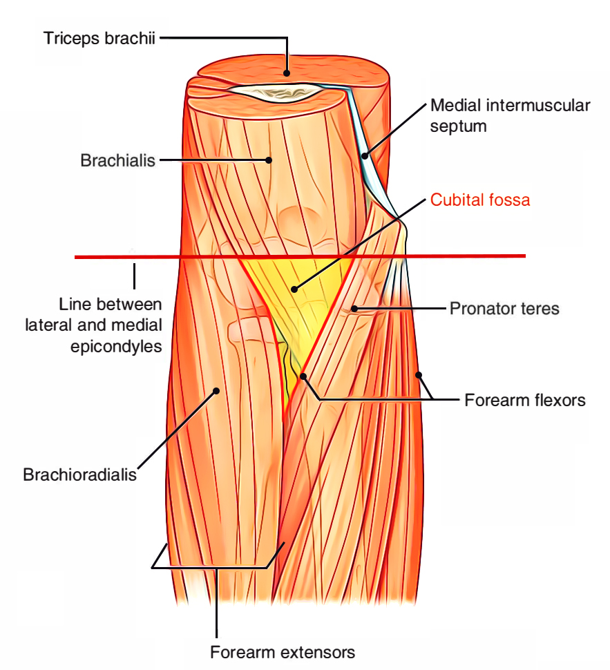
Cubital Fossa Margin
- Lateral: Medial border of brachioradialis muscle.
- Medial: Lateral border of pronator teres muscle.
- Base: An imaginary horizontal line, joining the front of 2 epicondyles of the humerus.
- Apex: Meeting point of the medial and lateral borders. Here brachioradialis overlaps the pronator teres.
- Floor: It’s created by 2 muscles, brachialis in the upper part and supinator in the lower part.
- Roof: It’s created from superficial to deep by:
- Skin
- Superficial fascia featuring (i) median cubital vein attaching cephalic and basilic veins, and (ii) medial and lateral cutaneous nerves of the forearm
- Deep fascia, reinforced by bicipital aponeurosis
Contents
The cubital fossa is really narrow space, and for that reason, its contents are shown only if the elbow is bent and its margins are pulled apart.
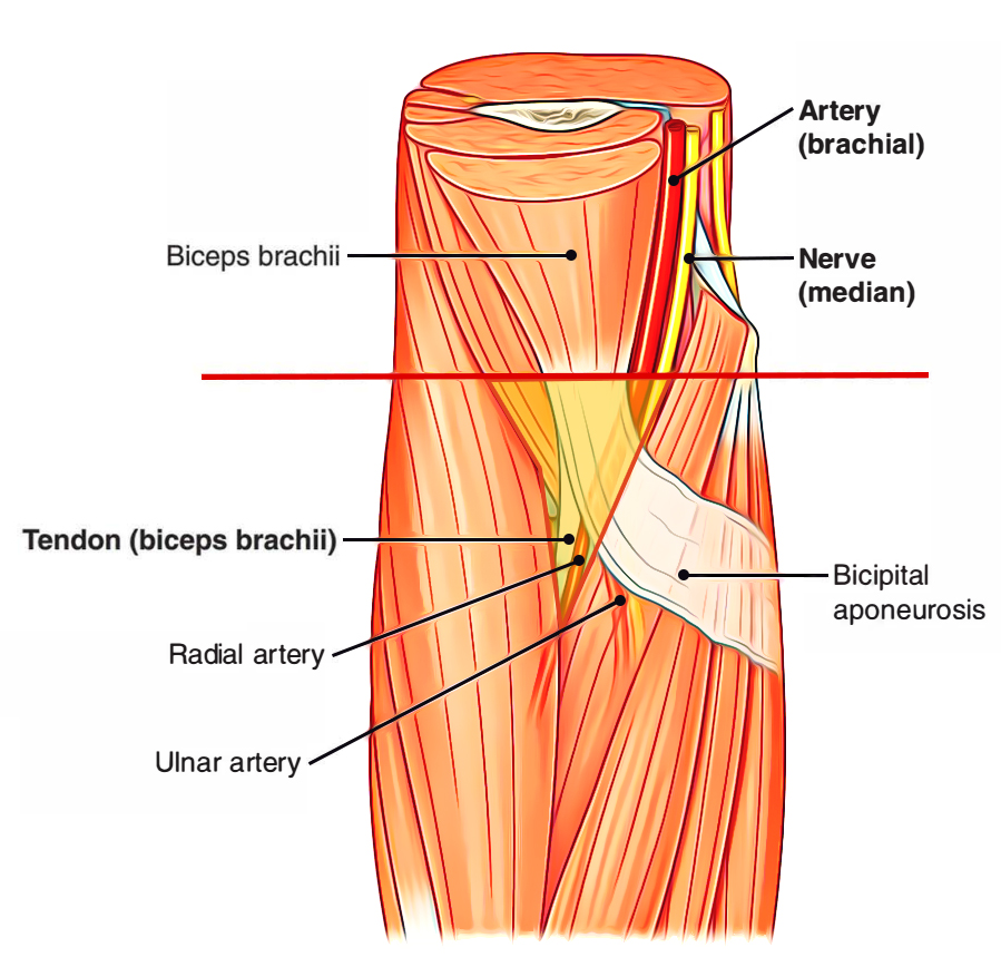
Cubital Fossa Content
The contents of cubital fossa from the medial to lateral side are as follows:
- Median nerve: It leaves the fossa by passing between 2 heads of pronator teres.
- Brachial artery: It ends in the fossa in the level of neck of radius by splitting into radial and ulnar arteries. The radial artery is superficial and makes the fossa in the apex. The ulnar artery is deep and enters deep to the pronator teres.
- Biceps tendon: It enters backwards and laterally to be connected on the radial tuberosity.
- Radial nerve: It is located in the gap between brachialis medially and brachioradialis laterally. At the level of lateral epicondyle it breaks up into 2 terminal branches: (a) superficial radial nerve and (b) deep radial nerve. The latter vanishes in the substance of supinator muscle. The superficial radial nerve enters downwards under the cover of brachioradialis.
Mnemonic: The contents of cubital fossa from the medial to lateral side are easily recalled by the mnemonic MBBS (M = Median nerve, B = Brachial artery, B = Biceps tendon, S = Superficial radial nerve
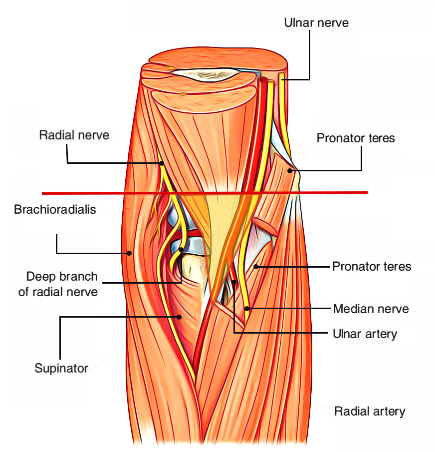
Cubital Fossa Position of Radial Nerve
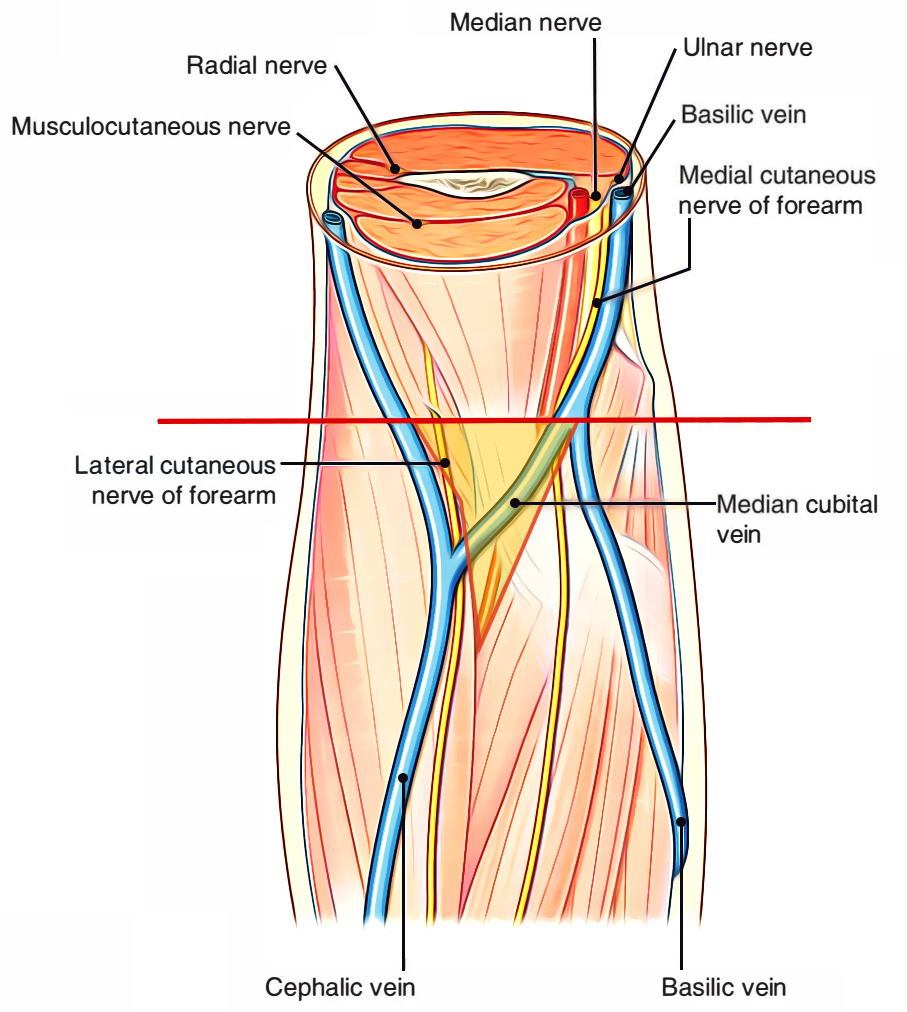
Cubital Fossa Superficial Structures
Clinical Relevance
The understanding of anatomy of the cubital fossa is medically essential for the following reasons:
- The median cubital vein in this region is the vein of selection for gathering blood samples and giving intravenous injections.
- The brachial pulse in this region is easily felt medial to biceps tendon, for recording the blood pressure.
To take care of the fractures around elbow, viz. supracondylar fracture of the humerus. The contents of cubital fossa notably the brachial artery and median nerve are exposed in supracondylar fracture of the humerus.

 (54 votes, average: 4.52 out of 5)