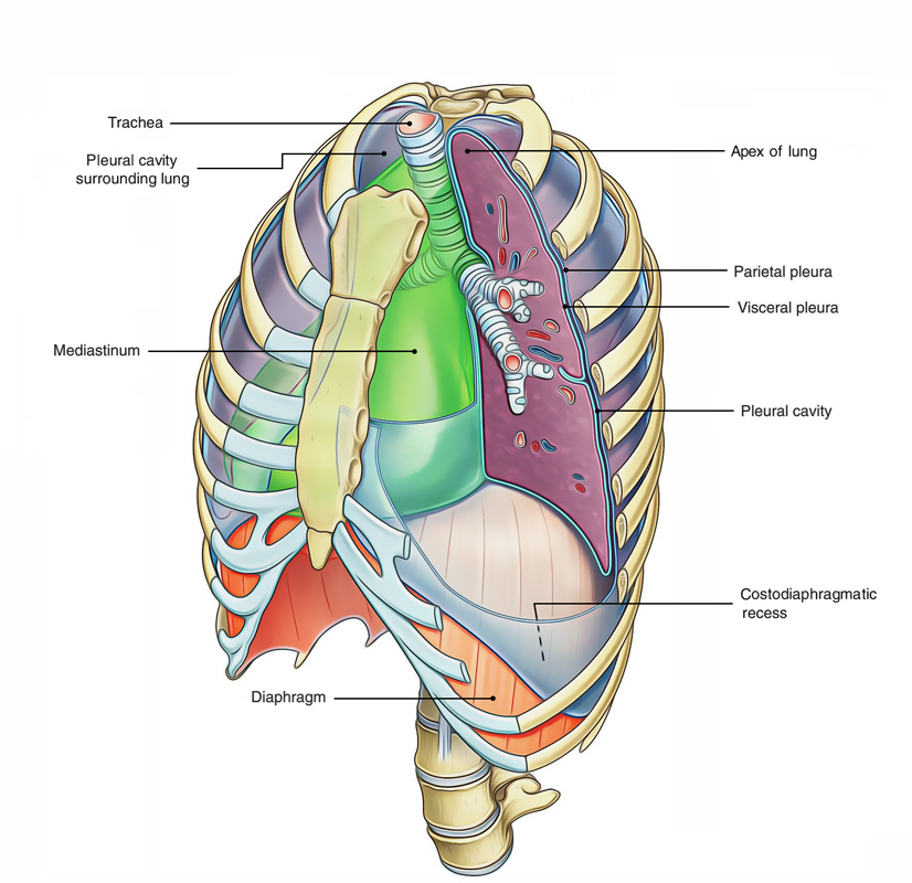
Every lung is invested by and enclosed in a serous sac which includes 2 constant serous membranes the visceral pleura and parietal pleura. The visceral pleura invests all the surfaces of the lung creating its gleaming outer surface, on the other hand the parietal pleura lines the pulmonary cavity (i.e., thoracic wall and mediastinum). The space between the visceral and parietal pleura is referred to as pleural cavity.

Pleural Cavities
A little description of development of lung makes it’s simpler to understand the relationship of the lung and pleura.
During early embryonic life, every lateral compartment of the thoracic cavity is inhabited by a closed serous sac, that is invaginated from the medial side by the growing lung and as an effect of the invagination, it converted into a double-layered sac. The outer layer is referred to as parietal pleura and the inner layer is termed visceral pleura. The visceral pleura is constant with parietal pleura in the root of the lung. The parietal and visceral layers are divided from every other by a slit-like potential space referred to as pleural cavity. The pleural cavity is normally filled up with a thin film of tissue fluid, which lubricates the adjoining surfaces of the pleura and enables them to move on every other without friction.
The 2 layers become constant with every other by way of a cuff of pleura, which encircles the root of the lung being composed of structures entering and leaving the lung at the hilum of the lung, like primary bronchi and root of the lung as far down as the diaphragm between the lung and the mediastinum.

 (60 votes, average: 4.78 out of 5)