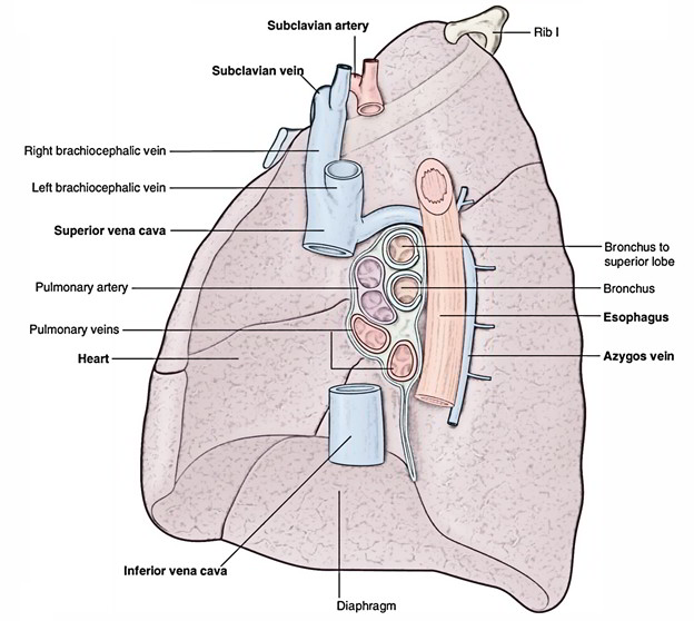
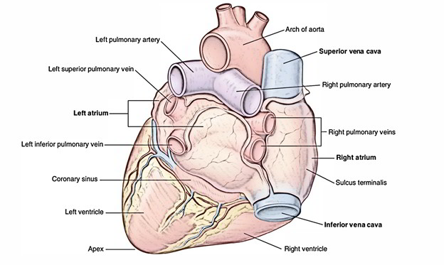
Pulmonary veins carry oxygenated blood via the lungs back to the left atrium of the heart. This makes the pulmonary veins different from the other veins in the body, which carry deoxygenated blood back to the heart from the rest of the body. On each side a Superior Pulmonary Vein and an Inferior Pulmonary Vein carry oxygenated blood from the lungs back to the heart. The veins arise at the Hilum of the Lung, travel via the Root of the Lung, and directly drain in the left atrium. Humans have four pulmonary veins, two from each lung.
Structure
Alveoli are tiny air sacs inside the lungs where oxygen and carbon dioxide are exchanged. These capillaries eventually join together to form a single blood vessel from each lobe of the lung. The pulmonary veins are constant with the capillary divisions of the pulmonary artery where they emerge via a capillary network on the edges of the alveoli. They create one vessel for each lung lobule by combining together. For each section these vessels combine as one forming a single large vein – three for the right lung and two for the left lung. Generally the vein from the middle lobe of the right lung and the one from the upper lobe merge with each other. Having two veins from the hilum of each lung is the most common pattern. Inside the posterior part of the left atrium they open up separately.

Pulmonary Veins
Circulation
There are normally four pulmonary veins, two supplying each lung:
- Right superior: The right upper and middle lobes.
- Right inferior: The right lower lobe.
- Left superior: The left upper lobe.
- Left inferior: The left lower lobe.
The pulmonary veins do not travel with the bronchi like the pulmonary arteries and they course in the intersegmental septa. The contact with deep bronchial veins inside the lung and with the superficial bronchial veins on the hilum is widespread.

Pulmonary Veins: Circulation
- Before taking a more vertical progression the inferior pulmonary veins travel parallel superficially and the superior pulmonary veins travel an oblique inferomedial route.
- They create a short intrapericardial section, to drain inside the left atrium when they travel via the lung hilum and antero-inferiorly to the pulmonary arteries.
- The ostia of the left pulmonary veins are more superior and the inferior pulmonary veins are more posteromedial.
Anatomical Variations
It not rare that the two left lobar veins end by a common opening into the left atrium and the three lobar veins on the right side remain separate rarely. As a result, in the healthy population the number of pulmonary veins attaching into the left atrium can vary between three and five. This creates variation in anatomy of many people that is observed in diagnosis.
Mutual Trunks
- Mutual draining trunk of left side – left superior and inferior pulmonary veins
Variant/Accessory/Additional Pulmonary Veins
- Single accessory: Right middle pulmonary vein which is around 10% in occurrence
- Two accessory: Right middle pulmonary veins
- One accessory: Right middle pulmonary vein and One accessory – Right superior pulmonary vein
- Superior section: Right inferior lobe vein
- Near base section: Right inferior lobe vein
- Right superior pulmonary vein that drains right superior basal segment
Clinical Significance
Total Anomalous Pulmonary Venous Connection
Generally, pulmonary veins return oxygenated blood to the left atrium via the lungs where it can then be circulated to the other parts of the body.
Total anomalous pulmonary venous connection a.k.a. total anomalous pulmonary venous drainage or total anomalous pulmonary venous return is a rare genetic cyanotic heart defect in which all four pulmonary veins create abnormal links to the systemic venous circulation and are located irregularly.
The disorder is lethal due to a deficiency of systemic blood flow if a patent foramen ovale, patent ductui arteriosa or an atrial septal defect is absent.

 (53 votes, average: 4.59 out of 5)