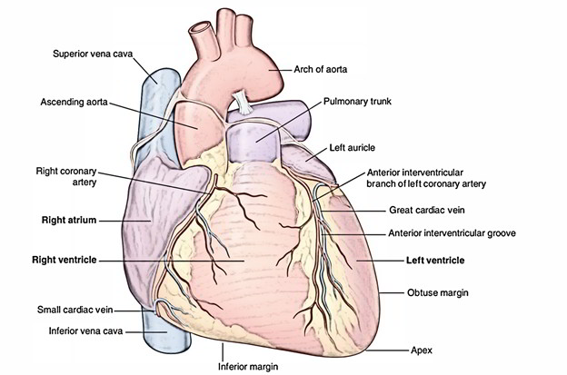
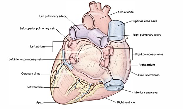
The pulmonary arteries emerge via the Truncus Arteriosus and the Sixth Pharyngeal Arch. It divides into two branches Right Pulmonary Artery and Left Pulmonary Artery. The right and left pulmonary arteries supply deoxygenated blood to the lungs via the right ventricle of the heart. The branching of the pulmonary trunk takes place anteroinferiorly towards the left of the branching of the trachea and near the left of the midline just inferior towards T4/T5 vertebral level. The Pulmonary arteries are the only arteries in the body that carry deoxygenated blood.

Pulmonary Arteries
Branches
Right Pulmonary Artery
The right pulmonary artery travels horizontally across the mediastinum and is longer compared to the left branch. It travels:
- Anteriorly towards the right main bronchus and anteriorly as well as a little inferiorly to the tracheal branching and,
- Posteriorly towards the ascending aorta, superior vena cava, along with upper right pulmonary vein.
The right pulmonary artery gives off a large branch to the superior lobe of the lung when it enters the root of the lung. The main vessel gives off a second (recurring) branch to the superior lobe, while it continues via the hilum of the lung and later divides to supply the middle as well as inferior lobes.
Left Pulmonary Artery
The left pulmonary artery is one of the branches of the pulmonary trunk that branches at the level of the transthoracic plane of Ludwig. It represents a direct posterior continuation of the pulmonary trunk and is shorter compared to the right pulmonary artery. It curves posterosuperiorly above the superior margin of the left main bronchus and also enters the superior part of the hilum of the left lung while posterior to the bronchus, where it divides in upper (ascending) and lower trunks.
- The Upper Lobar Artery is located medial to the left main bronchus and supplies the left Upper Lobe. This vessel may possibly emerge directly via the pulmonary artery or by the descending pulmonary artery.
- The Descending or Interlobar artery supplies the lingula of the left Upper Lobe and the Left Lower Lobe is located lateral to the left main bronchus.

Pulmonary Arteries: Branches
Development
The endocardial tubes create a swelling in the part near the heart around the third week of development of fetus. The swelling is called the Bulbus Cordis and the upper part of this swelling develops into the Truncus Arteriosus. The pulmonary arteries emerge via the Truncus Arteriosus and the sixth pharyngeal arch. The truncus arteriosis is created as a replacement to the Conus Arteriosus throughout the development of the heart. The structure has mesodermal origin.
During development of the heart, the truncus arteriosus is uncovered to what will ultimately be both of the left and right ventricles and then the heart tissues undergo folding. Two bulges are created on both sides of the truncus arteriosus as a septum is formed among the two ventricles of the heart. These expand gradually until the trunk divide into the aorta as well as pulmonary arteries.
Functions
- The pulmonary artery transport deoxygenated blood from the right ventricle to the lungs. The blood here becomes oxygenated as part of the process of respiration as it travels through capillaries surrounding the alveoli.
- The bronchial arteries supply nutrition to the lungs themselves as compared to the pulmonary arteries.
- The pulmonary artery pressure is an estimation of the blood pressure in the main pulmonary artery.
- This is measured by injecting a tube inside the main pulmonary artery. The average pressure is typically 9 – 18 mm Hg and the segment pressure measured in the left atrium is around 6-12mm Hg.
- In the cases of left heart failure, mitral valve stenosis, and other conditions, such as sickle cell disease, the wedge pressure may be elevated.
Clincal Significance
Pulmonary Hypertension
In a number of clinical conditions, the pulmonary artery has a major role. Pulmonary hypertension is used to describe a medical condition when there is an increase in the pressure of the pulmonary artery, when an average pulmonary artery pressure of greater than 25mmHg. An increased diameter of more than 29 mm diameter is generally used as a limiter to indicate pulmonary hypertension which can be measured on a CT scan. This maybe caused due to:
- Heart problems like heart failure.
- Lung or airway disease like COPD or scleroderma.
- Thromboembolic disease like pulmonary embolism or emboli observed in sickle cell anaemia.
Pulmonary Artery Stenosis
Pulmonary artery stenosis is a contraction (stenosis) that happens in the pulmonary artery. The narrowing may arise in the left or right pulmonary artery branches as well as/or in the main pulmonary artery. This narrowing makes it difficult for blood to reach the lungs to pick up oxygen. Without enough oxygen, the heart and body cannot function as they should. The right ventricle which pumps blood in the pulmonary arteries, in an effort to overpower the contraction increases the pressure in the chamber to levels that can be injurious to the heart muscle.

 (56 votes, average: 4.62 out of 5)