There are 2 superficial veins of the neck. All these are as follows:
- External jugular vein.
- Anterior jugular vein.
External Jugular Vein
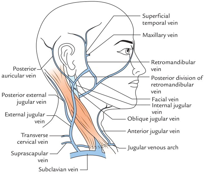
The starting point is just below the angle of the mandible by the joining of posterior section of retromandibular vein and posterior auricular vein. It then runs under the cover of platysma to pierce the deep cervical fascia in the anteroinferior angle of the posterior triangle about 2.5 cm above the clavicle along the posterior border of the sternocleidomastoid and enters the supraclavicular space, just about vertically downward across the sternocleidomastoid. It ends in the subclavian vein after going through this space. The external jugular vein has huge difference in its size and course. As it impedes the venous return to the right side of the heart and distends the vein, it becomes observable in old age especially when the individual holds his breath or blows his cheek with mouth closed.
Surface Anatomy
The external jugular vein can be marked on the surface by a line going downward and backward from angle of the mandible to the middle of the clavicle.
Its tributaries are as follows:
- Posterior auricular vein.
- Retromandibular vein.
- Posterior external jugular vein.
- Oblique jugular vein.
- Transverse cervical vein.
- Suprascapular vein.
- Anterior jugular vein.
Key Points
- The posterior auricular vein descends behind the auricle to join the posterior section of retromandibular vein. Posterior external jugular vein descends along the posterior border of sternocleidomastoid to join the external jugular vein a little below the midpoint of posterior border of the muscle. The oblique jugular vein interacts with all the internal jugular vein in the upper part of the neck. The suprascapular, transverse cervical and anterior jugular veins join the external jugular vein in the posterior triangle.
- There are just two pairs of valves in the lumen of the external jugular vein, one at its site of conclusion into the subclavian vein and the other about 4 cm above the clavicle. These valves, but don’t prevent regurgitation of blood.
Clinical Significance
The wall of the external jugular vein is adherent to deep fascia where it pierces the deep fascia (about 2.5 cm above the clavicle). For that reason, if external jugular vein is cut at this site, its walls can not fail. Hence, air is sucked into its lumen because of negative intrathoracic pressure during motivation resulting in venous air embolism that might cause passing later.
The external jugular vein is sometimes utilized for central venous cannulation, but because of its varying size and presence of valves it’s frequently hard to manipulate the cannula via the lower part of the vein. The right external jugular vein being in the most direct line together with the superior vena cava is the most generally utilized.
The external jugular vein is frequently examined by clinicians to measure the venous pressure in the right atrium. Normally, in recumbent (being located down) position, the lower one-third of the vein becomes filled up with blood but it fails on reclining at 45 ° angle. But if it stays complete even when the patient reclines at 45 ° angle, it proposes increased right atrial pressure frequently observed in congestive cardiac failure.
Anterior Jugular Vein
It starts below the chin in the submental region by the joining of small unnamed veins from the chin. It descends in the superficial fascia about 1 cm lateral to the midline. At about 2.5 cm above the suprasternal notch, it pierces the investing layer of deep cervical fascia to goes into the suprasternal space (of Burns), where it turns sharply laterally and enters deep to sternocleidomastoid and ends in the external jugular vein. In the suprasternal space, the anterior jugular vein is connected across the midline to its chap of opposite side by a transverse venous channel termed jugular venous arch.

 (57 votes, average: 4.82 out of 5)