The veins draining the upper limb, as elsewhere in the body, are split into 2 sets/groups:
- superficial
- deep
The superficial veins are found in the superficial fascia and are easily accessible. Being easily accessible, they’re frequently utilized by the clinicians for drawing blood samples or for giving intravenous injections.
The deep veins is located deep to muscles and accompany arteries as venae comitantes.
Superficial Veins
Superficial veins have the following general features:
- The superficial veins is located in the superficial fascia.
- The superficial veins have a tendency to create the pressure sites, for this reason they’re absent in the palm, along the ulnar border of the forearm, and back of the elbow.
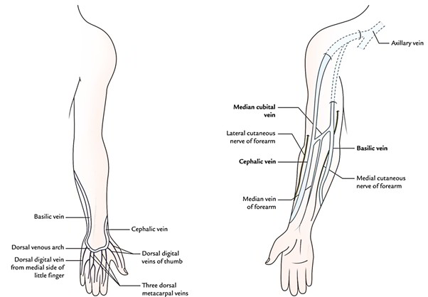
- There are 2 major superficial veins, one along the pre-axial border and the other along the post-axial border of the limb. The preaxial vein (cephalic vein) is longer compared to the postaxial vein (basilic vein), but the postaxial basilic vein drains more efficiently. The load of long cephalic vein is greatly relieved as a good amount of its blood is transferred to the efficient basilic vein by the median cubital vein (communicate channel).
The superficial veins are accompanied by the cutaneous nerves and superficial lymphatics.
Superficial veins comprise:
- Dorsal venous arch.
- Cephalic vein.
- Basilic vein.
- Median cubital vein.
Dorsal Venous Arch
The dorsal venous arch is a network of veins on the dorsum of hand. It presents irregular arrangement of veins generally with its transverse element, which is located 2-3 cm proximal to the heads of metatarsals.
Tributaries
The tributaries of dorsal venous arch are:
- 3 dorsal metacarpal veins.
- A dorsal digital vein from the medial side of little finger.
- A dorsal digital vein from the lateral side of index finger.
- 2 dorsal digital veins of the thumb.
- Veins draining palm of hand. These are (a) veins that circulate the margins of the hand and (b) perforating veins, which pass dorsally via the interosseous spaces.
- The dorsal venous arch drains into cephalic and basilic veins—the efferent vessels of dorsal venous arch.
The pressure on the palm during gripping doesn’t hamper the venous return of the palm, rather it facilities the return because venous blood from the palm is drained into dorsal venous arch.
Cephalic Vein
The cephalic vein begins as the continuation of lateral end of the dorsal venous arch.
It crosses the roof of anatomical box, ascends on the radial border of the forearm, continues upwards in front of elbow along the lateral border of biceps, pierces the deep fascia at the lower border of the pectoralis major, runs in cleft between the deltoid and pectoralis major (deltopectoral groove) up to the infraclavicular fossa, where it pierces the clavipectoral fascia and drains into the axillary vein.
Points to be noted
- At elbow, greater amount of blood from the cephalic vein is shunted in the basilic vein via median cubital vein.
- Cephalic vein is escorted by the lateral cutaneous nerve of the forearm.
- An accessory cephalic vein from back of the forearm (occasional) ends in the cephalic vein below the elbow.
- Cephalic vein is the preaxial vein of the upper limb and corresponds to the great saphenous vein of the lower limb.
Basilic Vein
The basilic vein begins as the continuation of the medial end of the dorsal venous arch of the hand. It runs upwards along the back of the medial border of the forearm, winds round this border near the elbow to reach the anterior aspect of the forearm, where it continues upwards in front of the elbow along the medial side of the biceps brachii up to the middle of the arm, where it pierces deep fascia, unites together with the brachial veins and runs along the medial side of the brachial artery to become continuous with the axillary vein at the lower border of the teres major.
- Basilic vein is the postaxial vein of the upper limb and corresponds to the short saphenous vein of the lower limb.
- About 2.5 cm above the medial epicondyle of humerus, it’s joined by the median cubital vein.
- It’s escorted by the medial cutaneous nerve of the forearm.
Median Cubital Vein
- It’s a communicating venous channel between the cephalic and basilic veins, which shunts blood from the cephalic vein to the basilic vein.
- It begins from the cephalic vein, 2.5 cm below the elbow bend, runs obliquely upwards and medially to end in the basilic vein, 2.5 cm above the bend of elbow.
The significant features of median cubital vein are as follows:
- It’s divided from brachial artery by the bicipital aponeurosis.
- It interacts with all the deep veins via a perforator vein, which pierces the bicipital aponeurosis.
- It gets median vein of the forearm.
- It shunts blood from cephalic vein to the basilic vein.
Median Vein of the Forearm
Median vein of the forearm begins from palmar venous network, runs upwards in the midline on the anterior aspect of forearm to end in any 1 of 3 veins in front of elbow (viz. cephalic, basilic, and median cubital veins).
Sometimes the upper end of median vein of the forearm bifurcates into median cephalic and median basilic veins, which join the cephalic and basilic veins, respectively. In this situation, the median cubital vein is absent.
Common Venous Patterns in front of the Elbow
The veins in front of the elbow commonly create 2 patterns, viz.
- H-shaped pattern.
- M-shaped pattern.
Clinical Significance
Venepuncture in the Cubital Fossa
The veins in front of the elbow, example, median cubital vein, cephalic vein, and basilic vein are routinely utilized for giving intravenous injections and for withdrawing blood from the donors. The median cubital vein is most preferred because of the following reasons:
1. It’s the most superficial vein in the body, for this reason access is easy.
2. It’s well supported by the underlying bicipital aponeurosis.
3. It’s well anchored to the deep vein by a perforating vein, for this reason it doesn’t slip during procedure.
- The cephalic vein is preferred for hemodialysis in the patients with chronic renal failure (CRF), to remove waste products from blood.
- The cut-down of cephalic vein in the deltopectoral groove is preferred when the superior vena cava infusion is essential.
- The basilic vein is preferred for cardiac catheterization for the following reasons:
- The diameter of basilic vein increases as it ascends from cubital fossa to the axillary vein.
- It’s in direct line together with the axillary vein. To goes into the right atrium the catheter enters in succession as follows:
Basilic vein —> axillary vein —> subclavian vein —> brachiocephalic vein —> superior vena cava —> right atrium of the heart.
The cephalic vein isn’t preferred for cardiac catheterization because of the following reasons:
- Its diameter doesn’t increase as it ascends.
- It joins the axillary vein at a right angle therefore it’s difficult to maneuver the catheter around sharp cephaloaxillary angle.
- In deltopectoral groove, it frequently divides into small branches. 1 of these branches ascends over the clavicle and joins the external jugular vein.
Deep Veins
The deep veins comprise:
- venae comitantes, which accompany the large arteries, like radial, ulnar, and brachial arteries.
- venae comitantes of the brachial artery.
- axillary vein.
Venae comitantes of the radial and ulnar arteries accompany the radial and ulnar arteries, respectively, and join to create the brachial veins.
Venae comitantes are small veins, 1 on every side of the brachial artery. They join axillary vein at the lower border of the teres major muscle. The medial one frequently joins the basilic vein.
Axillary vein begins as a continuation of basilic vein at the lower border of the teres major muscle and runs via axilla, goes through its apex to continue as subclavian vein in the outer border of the first rib.

 (47 votes, average: 4.64 out of 5)