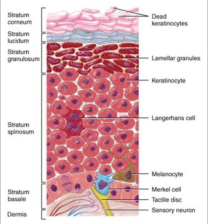
The epidermis is composed of five types of cells:
- Stem cells are undifferentiated cells that divide and give rise to the keratinocytes described next. They are found only in the deepest layer of the epidermis, called the stratum basale.
- Keratinocytes are the great majority of epidermal cells. They are named for their role in synthesizing keratin. In ordinary histological specimens, nearly all of the epidermal cells you see are keratinocytes.
- Melanocytes also occur only in the stratum basale, amid the stem cells and deepest keratinocytes. They synthesize the brown to black pigment melanin.
- They have branching processes that spread among the keratinocytes and continually shed melanin- containing fragments from their tips. The keratinocytes phagocytize these fragments and accumulate melanin granules on the “sunny side” of the nucleus. Like a parasol, the pigment shields the DNA from ultraviolet radiation.
- Tactile (Merkel) cells, relatively few in number, are receptors for the sense of touch. They, too, are found in the basal layer of the epidermis and are associated with an underlying dermal nerve fiber. The tactile cell and its nerve fiber are collectively called a tactile (Merkel) disc.
- Dendritic (Langerhans) cells are found in two layers of the epidermis called the stratum spinosum and stratum granulosum (described in the next section). They are macrophages that originate in the bone marrow but migrate to the epidermis and epithelia of the oral cavity, esophagus, and vagina. The epidermis has as many as 800 dendritic cells per square millimeter. They stand guard against toxins, microbes, and other pathogens that penetrate into the skin. When they detect such invaders, they alert the immune system so the body can defend itself.
Layers of the Epidermis
Cells of the epidermis are arranged in four to five zones, or strata (five in thick skin). The following description progresses from deep to superficial, and from the youngest to the oldest keratinocytes.
- The stratum basale (bah-SAY-lee) consists mainly of a single layer of cuboidal to low columnar stem cells and keratinocytes resting on the basement membrane. Scattered among these are the melanocytes and tactile cells. As stem cells of the stratum basale divide, they give rise to keratinocytes that migrate toward the skin surface and replace lost epidermal cells.
- The stratum spinosum (spy-NO-sum) consists of several layers of keratinocytes. In most skin, this is the thickest stratum, but in the thick skin it is usually exceeded by the stratum corneum. The deepest cells of the stratum spinosum remain capable of mitosis, but as they are pushed farther upward, they cease dividing. Instead, they produce more and more keratin filaments, which cause the cells to flatten. Therefore, the higher up you look in the stratum spinosum, the flatter the cells appear. Dendritic cells are also found throughout the stratum spinosum but are not usually visible in tissue sections.
The stratum spinosum is named for an artificial appearance (artifact) created by the histological fixation of tissue specimens. Keratinocytes are firmly attached to each other by numerous desmosomes, which partly account for the toughness of the epidermis. Histological fixatives shrink the keratinocytes so they pull away from each other, but they remain attached by the desmosomes—like two people holding hands while they step farther apart. The desmosomes thus create bridges from cell to cell, giving each cell a spiny appearance from which we derive the word spinosum. - Epidermal keratinocytes are also bound to each other by tight junctions, which make an essential contribution to water retention by the skin. The stratum granulosum consists of three to five layers of flat keratinocytes—more in the thick skin than in the thin skin. The keratinocytes of this layer contain coarse, dark-staining keratohyalin granules that give the layer its name. The functional significance of these granules will be explained shortly.
- The stratum lucidum (Loo-sih-dum) is a thin zone superficial to the stratum granulosum, seen only in the thick skin. Here, the keratinocytes are densely packed with a clear protein named eleidin (ee-LEE- ih-din). The cells have no nuclei or other organelles. This zone has a pale, featureless appearance with indistinct cell boundaries.
- The stratum corneum consists of up to 30 layers of dead, scaly, keratinized cells that form a durable surface layer. This layer is especially resistant to abrasion, penetration, and water loss.
Rate this Article: 
 (77 votes, average: 4.31 out of 5)
(77 votes, average: 4.31 out of 5)