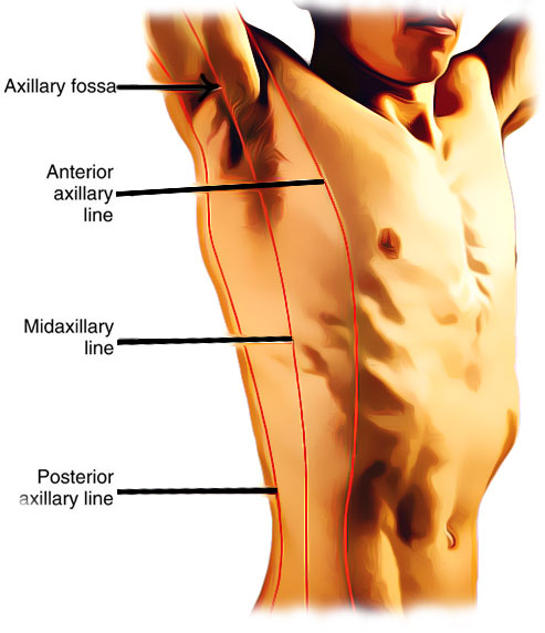
There are three axillary lines, anterior axillary line, midaxillary line and posterior axillary line.
Whenever there is abduction of the arm from the body by 90 degrees, by the anterior axillary fold, through the middle of the axilla, as well as from the posterior axillary fold imaginary vertical lines can be drawn (See the diagram below). These lines are respectively, referred to as the anterior axillary line, the midaxillary line, and the posterior axillary line.

Axillary Lines
The scapular line is a line drawn vertically from the superior and inferior poles of the scapula and the vertebral line is a line drawn down towards the center of the vertebral column.
Anterior Axillary Line
The anterior axillary line is the imaginary line that starts from the mid of the middle clavicle and the lateral end of the clavicle. It is on the anterior torso which is marked by the anterior axillary fold (which is formed by the lateral edge of the pectoralis major muscle).
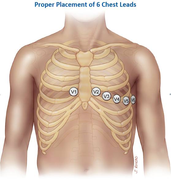
It is to be noted that the V5 ECG lead is placed on the anterior axillary line (see the image below).
Midaxillary Line
The midaxillary line is an imaginary line between the anterior axillary line and the posterior axillary line. It runs vertically down towards the surface of the body travelling through the tip of the axilla.
The anterior axillary line, which goes through the anterior axillary skinfold, and the posterior axillary line, which passes through the posterior axillary skinfold, are parallel to this line.
It is important to note that the midaxillary line is used in thoracentesis and V6 ECG lead is placed on this landmark.
Posterior Axillary Line
The posterior axillary line is an imaginary line on the posterior torso marked by the posterior axillary fold (which is a compound structure consisting of the latissimus dorsi and teres major muscles). It is drawn along the posterior axillary fold and followed down the chest wall.
It is to be noted that the V7 ECG lead is placed on the left posterior axillary line in the fifth intercostal space.


 (58 votes, average: 4.67 out of 5)