The Scapula (or shoulder blade) is a big, flattened, and triangular bone. The scapula is located on the upper part of the posterolateral aspect of the thorax, opposing second to seventh ribs.
A change in movement or scapular positioning might cause your shoulder, and can make it hard to move your arm when performing activities. It has two surfaces, three boundaries, three angles and three processes. When a condition or an injury causes these muscles to become imbalanced or weak, it can change the position of the scapula at rest or in motion.
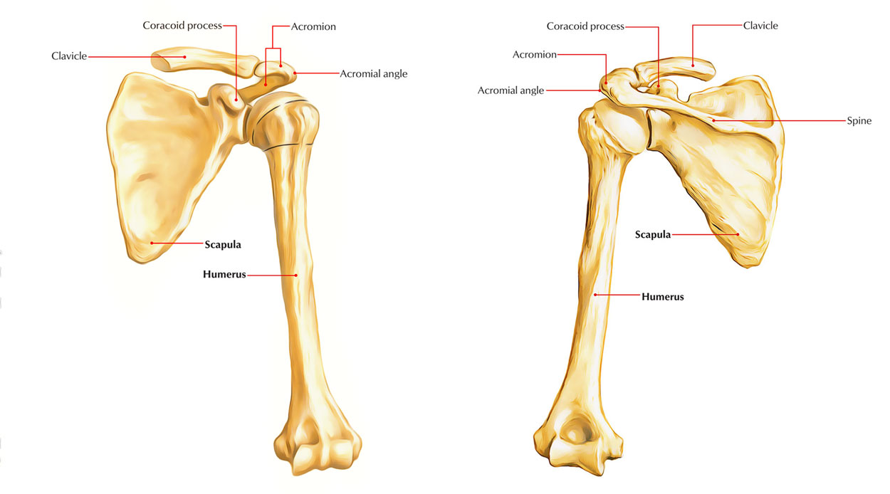
Scapula
Parts
The scapula is highly mobile and comprises of four parts : a body and three processes namely – spinous, acromion, and coracoid.
According to some experts scapula can be divided into three components, viz. head, neck, and body.
Body
The body is triangular, thin, and transparent. It has following features:
- Two surfaces: (a) costal and (b) dorsal.
- Three borders: (a) superior, (b) lateral, and (c) medial.
- Three angles: (a) inferior, (b) superior, and (c) lateral.
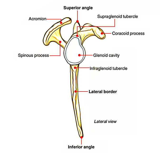
Scapula – Cross Section View
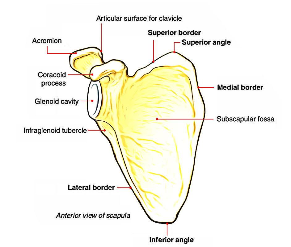
Parts of Scapula
The dorsal surface offers a shelf-like projection on its upper part named spinous process. The lateral angle is truncated to create an articular surface– the glenoid cavity. The lateral angle is thickened and referred to as head of the scapula, which is attached to the plate-like body by an inconspicuous neck.
Processes
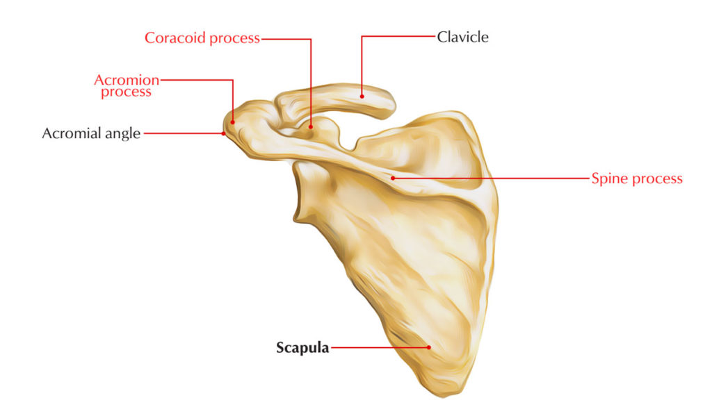
Scapula processes
There are three processes. These are as follows:
- Spinous process.
- Acromion process.
- Coracoid process.
The spinous process is a shelf-like bony projection on the dorsal aspect of the body. The acromion process projects forwards nearly at right angle from the lateral end of the spine. The coracoid process is like a birds beak. It starts from the upper border of the head and bends sharply to project superoanteriorly.
Anatomical Position And Side Determination
The side of the scapula can be figured out by holding the scapula in such a way that:
- The glenoid cavity faces laterally, forwards, and a bit upwards (at an angle of 45 ° from the coronal plane).
- The coracoid process is directed forwards.
- The shelf-like spinous process is directed posteriorly.
Features And Attachments
Surfaces
Costal Surface (Subscapular Fossa)
- It is concave and directed medially and forwards.
- It supplies three longitudinal ridges, which in turn supply connection to the intramuscular tendons of subscapularis muscle.
- The subscapularis muscle (a multipennate muscle) arises from the medial two-third of subscapular fossa/ costal surface with the exception of near the neck where a subscapular bursa intervenes in between the neck and the subscapular tendon.
- The serratus anterior muscle is inserted on this surface with the medial border and inferior angle.
Dorsal Surface
- The dorsal surface is convex and presents a shelf-like projection referred to as spinous process.
- The spinous process divides the dorsal surface into supraspinous and infraspinous fossae. The upper, supraspinous fossa is smaller (one-third) and lower, infraspinous fossa is larger (two-third).
- The spinoglenoid notch is located between lateral border of the spinous process and the dorsal surface of the neck of scapula. By means of this notch supraspinous fossa interacts with the infraspinous fossa and suprascapular nerve and vessels pass from supraspinous fossa to the infraspinous fossa.
- The supraspinatus muscle starts from medial two-third of supraspinous fossa.
- The infraspinatus muscle starts from medial two-third of infraspinous fossa.
- The teres minor muscle starts from the upper two-third of the dorsal surface of lateral border. This origin is interrupted by circumflex scapular artery.
- The teres major muscle arises from the lower one-third of the dorsal surface of lateral border and inferior angle of scapula.
- The latissimus dorsi muscle also starts from dorsal surface of the inferior angle by a small slip.
Borders
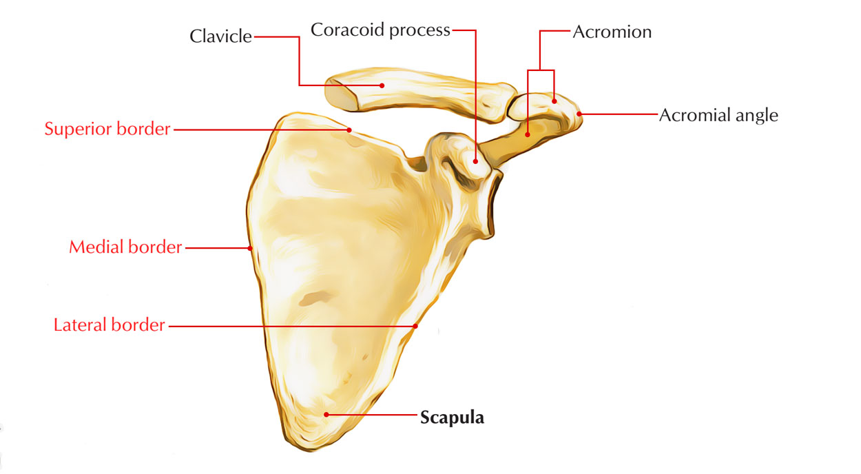
Scapula Borders
Superior Border
- The superior border is the shortest border and extends between superior and lateral angles.
- The suprascapular notch exists on this border near the root of coracoid process.
- The suprascapular notch is transformed into suprascapular foramen by superior transverse (suprascapular) ligament.
- The suprascapular artery passes above the ligament and suprascapular nerve passes below the ligament, via suprascapular foramen. (Mnemonic: Air force flies above the Navy, i.e., A: artery is above and N: nerve is below the ligament.)
- The inferior belly of omohyoid arises from the superior border near the suprascapular notch.
Lateral Border
- The lateral border is the thickest border and extends from inferior angle to the glenoid cavity.
- The infraglenoid tubercle is present at its upper end, just below the glenoid cavity.
- The long head of triceps muscle starts from the infraglenoid tubercle.
Lateral border of scapula is thick due to the fact that it serves as fulcrum during rotation of the scapula.
Medial Border (Vertebral Border)
- It goes on from superior angle to the inferior angle.
- It is thin and angled at the root of spine of scapula.
- The serratus anterior muscle is inserted on the costal surface of the medial border and the inferior angle.
- The levator scapulae muscle is inserted on the dorsal aspect of the medial border from superior angle to the root of spine.
- The rhomboideus minor muscle is inserted on the dorsal aspect of the medial border opposite the root of spine.
- The rhomboideus major muscle is inserted on the dorsal aspect of the medial border from the root of spine to the inferior angle.
Angles
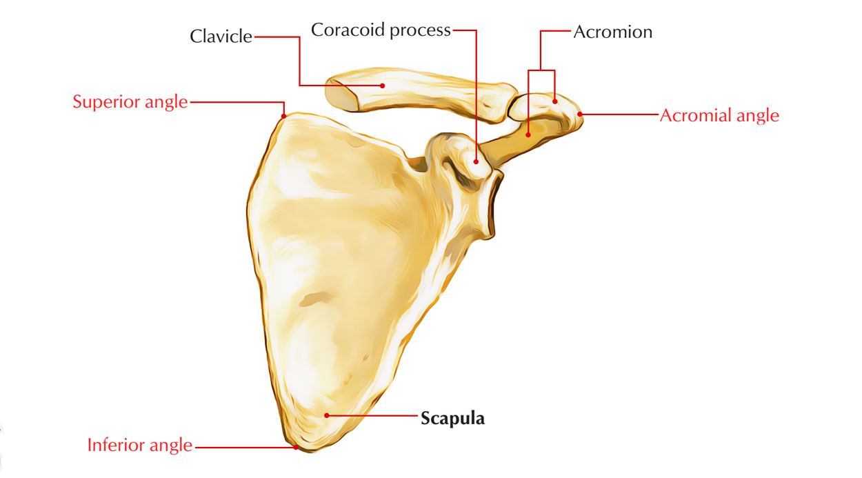
Scapula angles
Inferior angle:
- It is located over the 7th rib or the 7th intercostal space.
Superior angle:
- It is at the junction of superior and medial borders, and is located over the 2nd rib.
Lateral angle (head of scapula)
- It is truncated and bears a pear-shaped articular cavity referred to as the glenoid cavity, which articulates along with the head of humerus to form glenohumeral (shoulder) joint.
- A fibrocartilaginous rim, the glenoid labrum is connected to the margins of glenoid cavity to deepen its concavity.
- The capsule of shoulder joint is connected to the margins of glenoid cavity, proximal to the attachment of glenoid labrum.
- The long head of biceps brachii emerges from supr aglenoid tubercle. This origin is intracapsular.
Processes
Spinous Process (Spine Of Scapula)
- It is a triangular shelf-like bony projection, connected to the dorsal surface of scapula at the junction of its upper one-third and lower two-third.
- It divides the dorsal surface of scapula into two parts– upper supraspinous fossa and lower infraspinous fossa.
- The spine has two surfaces–(a) superior and (b) inferior, and three borders–(a) anterior, (b) posterior, and (c) lateral.
Surfaces
- The superior surface of spine forms the lower boundary of supraspinous fossa and gives origin to supraspinatus.
- The inferior surface of spine forms the upper limit of infraspinous fossa and gives origin to infraspinatus.
Borders
- The anterior border of spine is connected to the dorsal surface of scapula.
- The lateral border of spine bounds the spinoglenoid notch through which pass suprascapular nerve and vessels from supraspinous fossa to infraspinous fossa.
- The posterior border of spine is also named crest of spine. Trapezius is inserted to the upper lip of crest of spine, while posterior fibres of deltoid take origin from its lower lip.
Acromion Process (Acromion)
- It projects forwards nearly at right angle from the lateral end of spine and overhangs the glenoid cavity.
- Its superior surface is subcutaneous.
- It has a tip, two borders (medial and lateral), and two surfaces (superior and inferior).
- The medial and lateral borders of acromion continue with the upper and lower lips of the crest of the spine of scapula, respectively.
- Its superior surface is rough and subcutaneous.
- Its inferior surface is smooth and related to subacromial bursa.
- The medial border of acromion presents insertion to the trapezius muscle. Close to the tip, medial border presents a circular facet, which articulates with the lateral end of clavicle to form the acromioclavicular joint.
- The lateral border of acromion gives origin to intermediate fibres of the deltoid muscle.
- The coracoacromial ligament is connected to the tip of acromion.
- The acromial angle is found at the junction of lateral border of acromion and lateral border of the crest of the spine of scapula.
Coracoid Process
- It starts from the upper part of the head of scapula and bent sharply so as to project forwards and slightly laterally.
- The coracoid process gives attachment to three muscles— short head of biceps brachii, coracobrachialis, and pectoralis minor, and three ligaments – coracoacromial, coracoclavicular, and coracohumeral.
- The short head of biceps brachii and coracobrachialis arise from its tip by a common tendon.
- The pectoralis minor muscle is inserted on the medial border of the upper surface.
- The coracoacromial ligament is connected to its lateral border.
- The conoid part of the coracoclavicular ligament (rhomboid ligament) is connected to its knuckle.
- The trapezoid part of the coracoclavicular ligament (rhomboid ligament) is connected to a ridge on its superior aspect between the pectoralis minor muscle and coracoacromial ligament.
- The coracohumeral ligament is connected to its root adjacent to the glenoid cavity.
Ossification
The ossification of scapula is cartilaginous. The cartilaginous scapula is ossified by eight centres— one primary and seven secondary. The primary centre appears in the body. The secondary centres appear as follows:
- Two centres appear in the coracoid process.
- Two centres appear in the acromion process.
- One centre shows up each in the (a) medial border, (b) inferior angle, and (c) in the lower component of the rim of glenoid cavity.
The primary centre in the body and first secondary centre in the coracoid process shows up in eighth week of intrauterine life (IUL) and first year of postnatal life, respectively and they merge at the age of 15 years.
All other secondary centres show up at about puberty and merge by 20th year.
Clinical Significance
Sprengel’s deformity of the scapula (congenital high scapula): The scapula forms in the neck region during intrauterine life and after that it migrates downwards to its adult position (i.e., upper part of the back of the chest). Failure of descent causes Sprengel’s deformity of the scapula. In this condition the scapula is hypoplastic and located in the neck region. It may be connected to the cervical part of vertebral column by a fibrous, cartilaginous, or bony bar referred to as omovertebral body. An attempt to pull down scapula by a surgical procedure may cause injury to the brachial plexus.
- In living individual, the tip of coracoid process can possibly be palpated 2.5 cm below the junction of lateral one-fourth and medial three-fourth of the clavicle.
- In reptiles, coracoid process is a separate bone, but in humans it is connected to scapula and therefore it represents atavistic epiphysis.
- 1st coracoid centre represents precoracoid element and 2nd coracoid (subcoracoid) centre represents coracoid proper of reptilian girdle.

 (62 votes, average: 4.85 out of 5)