The muscles of mastication are concerned with movements of mandible in the temporomandibular joints during mastication.
They’re split into 2 groups:
Main muscles:
- Temporalis.
- Masseter.
- Lateral pterygoid.
- Medial pterygoid.
Accessory muscles:
- Digastric.
- Buccinator.
- Mylohyoid.
- Geniohyoid.
Primary Muscles of Mastication
The characteristic features of the main muscles of mastication are as follows:
- All are found in or around the infratemporal fossa.
- All are added into the ramus of the mandible.
- All are innervated by the mandibular division of the trigeminal nerve.
- All are concerned with movements of the mandible on the temporomandibular joints.
- All grow from mesoderm of the very first pharyngeal arch.
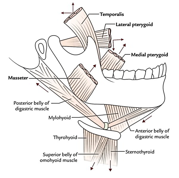
Temporalis
It’s connected above to the temporal line and below to the zygomatic arch and is covered by tough temporal fascia. It’s located in the temporal fossa and is a fan-shaped muscle.
Origin
It appears from:
- Whole of the floor of temporal fossa with the exception of the part created by the zygomatic bone.
- Deep surface of the temporal fascia.
Insertion
The fibres converge and descend to create a tendon, which goes through the gap between the zygomatic arch and the side of the skull. The muscle is added into:
- The medial surface, apex, anterior, border of the coronoid process of ramus of mandible
- The anterior border of the ramus of mandible, nearly up to the final molar tooth
The temporalis muscle is fan shaped. The anterior fibres are oriented vertically, the posteriormost fibres are disposed nearly horizontally and interceding intermediate fibres are positioned obliquely.
Nerve Supply
The temporalis is supplied by the anterior and posterior deep temporal nerves, the branches of the anterior section of the mandibular nerve.
Activities
The temporalis muscle elevates the mandible and so shuts the mouth and approximates the teeth. This movement needs both the upward pull of the anterior fibres and backward pull of the posterior fibres.
Posterior fibres retract the mandible after it’s been protruded.
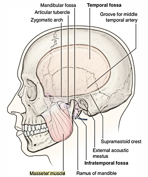
Masseter
The masseter (Greek: masseter = a chewer) is a thick quadrilateral muscle covering the lateral surface of the ramus of the mandible consisting of its coronoid process. The condylar process is left uncovered.
Origin
The masseter is composed of the following 3 layers:
- Superficial layer.
- Middle layer.
- Deep layer.
Superficial layer is largest of the 3 layers of masseter and originates by a thick aponeurosis from: maxillary process of zygomatic bone and anterior two-third of the inferior border of the zygomatic arch.
Middle layer originates from lower border of the posterior one-third of the zygomatic arch.
Deep layer originates from deep surface of the zygomatic arch.
Insertion
Superficial fibres pass downwards and backwards at 45° to be added into the angle and lower posterior half of the lateral surface of the ramus of the mandible.
Middle fibres pass vertically downwards to add into the central part of the ramus.
Deep fibres pass vertically downwards to fit into the upper part of the mandibular ramus and its coronoid process.
Key Points
Intramuscular tendinous septa in the superficial layer are liable for creating ridges on the ramus of the mandible.
Middle and deep fibres collectively represent the deep part of the masseter.
Nerve Supply
The masseter is supplied by a masseteric nerve, a branch from anterior section of the mandibular nerve.
Activities
The masseter muscle elevates the mandible to shut the mouth.
Lateral Pterygoid
It’s a short, thick conical muscle with its apex pointing backwards. It enters backwards and somewhat laterally from the roofing and medial wall of the fossa to the neck of the mandible.
Origin
The lateral pterygoid is composed of 2 heads, upper and lower:
- The upper smaller head appears from the temple surface and crest of the higher wing of the sphenoid bone.
- The lower bigger head originates from the lateral surface of the lateral pterygoid plate of the sphenoid bone.
Insertion
The fibres of 2 heads run backwards and laterally and converge to create a thick tendon that is added into:
- Pterygoid fovea on the very front of the neck of the mandible.
- Articular disc and capsule of the temporomandibular joint.
Nerve Supply
Lateral pterygoid is supplied by a branch of anterior section of the mandibular nerve.
Activities
- Lateral pterygoids of 2 sides depress the mandible (opens the mouth) by pulling forwards the condylar processes of the mandible and the articular discs of the temporomandibular joints.
- Medial and lateral Pterygoid muscles of 2 sides acting jointly protrude the mandible.
- Medial and lateral pterygoid muscles of the 2 sides contract alternately to generate side to side movements of the lower jaw as in mastication.
Things to Remember
- The lower head of lateral pterygoid enters between the 2 heads of the medial pterygoid muscle.
- It’s the only masticatory muscle, which opens the mouth.
- The articular disc of temporomandibular joint is developmentally a part of tendon of lateral pterygoid muscle.
Connections
The lateral pterygoid is regarded as the key muscle of theinfratemporal region because its connections supply a rational ideaabout the layout of structures in this region. Its connections are:
Superficial:
- Ramus of the mandible
- Masseter
- Tendon of temporalis
- Superficial head of medial pterygoid
- Maxillary artery and its temporal and masseteric branches
Deep:
- Mandibular nerve
- Middle meningeal artery
- Sphenomandibular ligament
- Deep head of medial pterygoid muscle
Structures appearing at the upper border:
- Deep temporal nerves (2 in number)
- Masseteric nerve
Structures appearing at the lower border:
- Inferior alveolar nerve and artery
- Lingual nerve
- Middle meningeal artery (it enters up deep to the lower border)
Structures going through the gap between the 2 heads:
- Maxillary artery, which enters the gap to get to the pterygopalatine fossa via pterygomaxillary fissure
- Buccal nerve, a branch of mandibular nerve.
- It comes out via the gap to supply sensory innervation to the skin and mucus membrane of the cheek.
Medial Pterygoid
The medial pterygoid is a thick quadrilateral muscle and is composed of 2 heads: superficial and deep.
Origin
- The small superficial head (a small slide of muscle) originates from maxillary tuberosity and lateral surface of the pyramidal process of palatine bone.
- The large deep head (creating the majority of muscle) appears from medial surface of the lateral pterygoid plate and grooved surface of the pyramidal process of palatine bone.
Insertion
The fibres run downwards, backwards and laterally to be added by a powerful tendinous lamina into a roughened area on the posteroinferior part of the medial surface and angle of ramus of mandible as high as the mandibular foramen and as forwards as the mylohyoid groove.
Nerve Supply
The medial pterygoid is supplied by a nerve to medial pterygoid, a branch from the primary trunk of the mandibular nerve.
Connections
Superficial:
- Lingual nerve
- Inferior alveolar nerve
- Inferior alveolar vessels
Deep:
- Levator palati and tensor palati muscles
- Superior constrictor of pharynx
- Styloglossus and stylopharyngeus muscles.
Activities
- Medial pterygoids of 2 sides elevate the mandible to assist in closure of mouth.
- Acting with lateral pterygoids, the medial pterygoids protrude the mandible.
- When medial and lateral pterygoids of 1 side act collectively, the corresponding side of the mandible is rotated forwards and to the opposite side.
- Medial and lateral pterygoids of 2 sides when contract alternately generate side to side movements that are utilized to grind the food.
Origin, Insertion, Nerve Supply and Activities of Main Muscles of Mastication
| Muscles | Origin | Insertion | Nerve supply | Actions |
|---|---|---|---|---|
| Temporalis (fan shaped) | Floor of temporal fossa Temporal fascia | Tip, anterior border, and medial surface of coronoid process Anterior border of ramus of mandible | Mandibular division of trigeminal nerve | Elevation of mandible by anterior and middle fibres Retraction of mandible by posterior fibres |
| Masseter (quadrilateral) | Zygomatic arch adjoining part ofzygomatic process of maxilla | Lateral surface of ramus of mandible Coronoid process | Mandibular division of trigeminal nerve | Elevation of mandible to occlude the teeth for forceful bite |
| Lateral pterygoid (Short, thick conical) | Upper head from infratemporal surface and crest of greater wing of sphenoid. Lower head from lateral surface of lateral pterygoid plate | Pterygoid fovea on anterior surface of neck of mandible Articular disc and capsule of TMJ | Mandibular division of trigeminal nerve | Depression of mandible by pulling the neck of mandible forward Protraction |
| Medial pterygoid (quadrilateral) | Superficial head from tuberosity of maxilla Deep head from medial surface of lateral pterygoid plate | Medial surface of angle adjoining ramus of mandible | Mandibular division of trigeminal nerve | Elevation of mandible Protraction |
The main muscles of mastication in many cases are referred to as muscles of mastication.
Clinical Significance
The muscles of mastication and their motor innervation can be analyzed medically by requesting the patient to clench his teeth repeatedly and after that palpating the temporalis and masseter in the temporal fossa and over the ramus of mandible, respectively.
Test Your Knowledge
Muscles of Mastication

 (55 votes, average: 4.63 out of 5)