The Patella is the largest sesamoid bone, seen in the tendon of quadriceps femoris. It’s situated in front of the knee joint, thus it’s also termed knee cap. It’s a flattened and triangular bone with all the base facing upward, and the apex downward. Its anterior aspect is convex and rough, on the other hand its posterior surface presents a large articular surface split into small medial part and large lateral part.

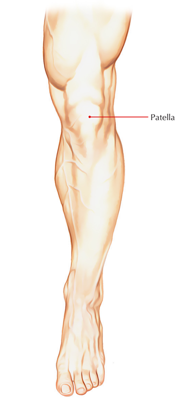
Patella
Anatomical Position and Side Decision
Hold the patella in this style that:
- Its apex faces downward and its base faces upward.
- Its articular surface faces posteriorly. The large lateral part of articular surface determines the side.
- Trick apparatus: Keep the articular surface of patella on the table-top in this manner that its base is directed toward you and its apex far from you. Now find the tilt of patella. The patella, usually, constantly tilts toward the side it belongs to.
Features and Attachments

Patella: Features and Attachments
The patella presents these features:
- Apex.
- 3 edges: Superior, medial, and lateral.
- 2 surfaces: Anterior and posterior.
Apex
It’s directed downward and gives connection to the ligamentum patellae, which attaches the patella to the tibial tubercle.
Borders
Superior Border

Patella: Borders
The superior border is also named as the base. It gives connection to rectus femoris in the anterior part and vastus intermedius in its posterior part.
Lateral Border
- It gives connection to vastus lateralis in its upper one-third and lateral patellar retinaculum in the lower two-third.
Medial Border
- It gives connection to vastus medialis in the upper two-third and medial patellar retinaculum in its lower one-third.
The medial and lateral patellar retinaculum are the growths of the tendons of vastus lateralis and vastus medialis, respectively.
Surfaces
Anterior Surface
- It’s rough, convex, longitudinally striated, and presents numerous vascular foramina.
- It’s subcutaneous and subcutaneous prepatellar bursaintervenes between it and skin.
Posterior Surface
1. Its lower quarter is rough and non-articular, while its upper three-fourth is smooth and articular.
2. Small non-articular part near the apex is split into 2 regions: lower and upper.
- Lower area gives connection to ligamentum patellae.
- Upper area is related to infrapatellar pad of fat.
3. The large articular area is mainly split by a vertical ridge into a bigger lateral and a smaller medial area as a way to fit with the mutual articular surfaces of the femur. The vertical ridge itself takes up the groove on the patellar surface of femoral lower end in the position of expansion.
- Larger lateral articular area is located in contact together with the lateral femoral condyle in all places of the knee.
- Small medial articular area is further split by another vertical ridge into a large lateral part and a narrow medial part referred to as medial strip. The medial strip comes in touch with the under surface of medial femoral condyle during total flexion in the knee.
Making apart this medial strip, entire of articular outermost layer of the patella is subdivided into upper, middle, and lower regions by 2 faint horizontal lines.
The patellar surface of femur comes in touch together with the upper area in common flexion, together with the middle area in semiflexion, and with the lower area in average extension.
Clinical Significance
Dislocation of The Patella
The patella has natural inclination to dislocate laterally because of upward and lateral pull by the quadriceps. Nevertheless, it’s countered by 3 variables:
- More forward projection of lateral femoral condyle.
- More protracted insertion of vastus medialis to the medial border of the patella than that of vastus lateralis on the lateral border.
- Medial pull used by medial patellar retinaculum.
Fracture of The Patella
- (a) A direct blow on the patella fractures it into 2 or more bits
- (b) a sudden and strong contraction of quadriceps femoris causes a transverse fracture of the patella. The patella being a sesamoid bone is devoid of the periosteum, thus when fractured bony union doesn’t take place.
Ossification
The patella ossifies from several centers which appear during 3-6 years and fuse immediately to create an individual center. The ossification of patella is finished at puberty.
Clinical Significance of ossification
Abnormal ossification of the patella. Abnormal ossification centers in the patella may create 2 or 3 different sections of the patella (referred to as bipartite or tripartite patella). This state can be discerned from fracture of the patella as it’s bilateral and the pieces have smooth margins.
Test Your Knowledge
Patella

 (52 votes, average: 4.67 out of 5)