The Posterior Compartment of Thigh is the gluteal fold above to the rear of the knee below.
Cutaneous Innervation
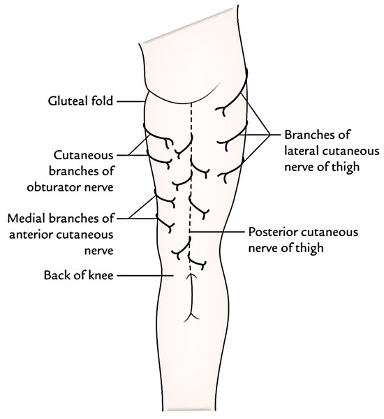
The nervous supply of skin of the back of the thigh is by following nerves
- Posterior cutaneous nerve of thigh goes superficially to long head of bicep femoris and downwards in the mid-section line on back of thigh deep to fascia lata. It follows the short saphenous vein into middle of calf and cleaves through the fascial roof of the fossa on reaching the popliteal fossa. Its branches supply the proximal part of the back of the leg and the skin on the back of the thigh.
- The medial part of the back thigh in the upper part is supplied by Cutaneous branches of obturator nerve.
- Medial part of the back of the thigh in the lower part is supplied by medial branches of the anterior cutaneous nerve.
- In the posterior part of thigh in the upper part has small branches sent by Lateral cutaneous nerve.
Contents of Posterior Compartment of the Thigh
The posterior compartment of the thigh is also named flexor compartment. It’s totally divided from the anterior compartment by the lateral intermuscular septum, but it’s incompletely divided from the medial compartment of the thigh by the ill-defined posterior intermuscular septum.
The contents of the posterior compartment of the thigh are as follows:
- Muscles: Hamstring muscles and short head of the biceps femoris.
- Nerve: Sciatic nerve.
- Arteries: Arterial anastomoses on the back of the thigh.
Hamstring (Muscles on the Back of the thigh)
These muscles on the back of the thigh are termed the hamstring muscles (Latin ham = back of thigh). Yet conventionally the short head of the biceps femoris isn’t incorporated in the hamstring group.
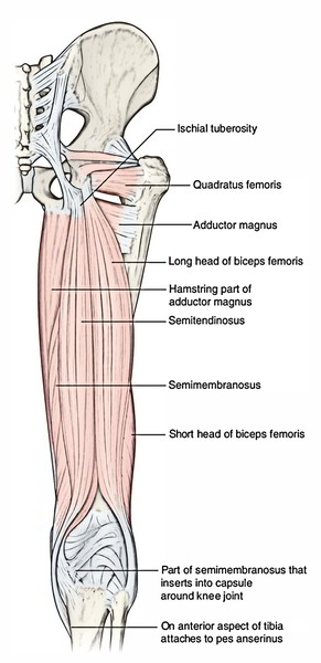
The hamstring muscles are:
- Semitendinosus.
- Semimembranosus.
- Biceps femoris (long head).
- Ischial head of adductor magnus.
The characteristic features of hamstring muscles are:
- All originate from the ischial tuberosity.
- All are added into one of the bones of the leg.
- All are furnished by tibial part of the sciatic nerve.
- All are flexors of the knee and extensors of the hip joint.
Points to be noticed
- The posterior thigh muscles were called “hamstrings” because their tendons on the rear of knee are accustomed to hang up hams (hip and thigh regions of critters viz., pigs.).
- The adductor magnus reaches only up to the adductor tubercle of the femur, but is comprised amongst the hamstrings group of muscles since the tibial collateral ligament of the knee joint morphologically represents the degenerated tendon of the muscle, that is connected below on the tibia.
Origin, Insertion, And Nerve Supply of The Muscles On The Back of thigh
| Muscle | Origin | Insertion | Nerve supply |
|---|---|---|---|
| Biceps femoris (a) Long head | (a) Long head: From lower medial part of upper quadrilateral area of ischial tuberosity | Into the head of the fibula in front of its styloid process | (a) Long head, by the tibial part of the sciatic nerve (L5; S1, S2) |
| (b) Short head | (b) Short head: From lateral lip of the linea aspera and from the upper two-third of the lateral supracondylar line | (b) Short head by the common peroneal part of the sciatic nerve (L5; S1, S2) | |
| Semitendinosus | From the lower medial part of upper quadrilateral area of the ischial tuberosity, | Into the upper part of the medial surface of the tibia | Tibial part of the sciatic nerve (L5; S1, S2) |
| Semimembranosus | From the upper lateral part of upper quadrilateral area of ischial tuberosity | Into the horizontal groove on the posterior aspect of the medial condyle of the tibia | Tibial part of the sciatic nerve (L5; S1, S2) |
| Ischial part of adductor magnus | From inferolateral aspect of the ischial tuberosity | Into the adductor tubercle | Tibial part of the sciatic nerve (L5; S1, S2) |
See Hamstring for more details.
Sciatic Nerve
The sciatic nerve is the thickest nerve within the body. It’s around 1.5 to 2 cm wide at the start. It originates from sacral plexus in the pelvis and is composed of 2 parts- tibial part and common peroneal part.
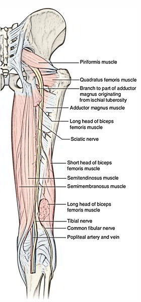
The tibial part is composed by the ventral sections of anterior primary rami of L4, L5; S1, S2, S3.
The common part is composed by the dorsal sections of anterior primary rami of L4, L5; S1, S2.
The 2 parts are typically enclosed in a standard sheath of the connective tissue.
See Sciatic Nerve for more detail.
Arterial Anastomoses on the Back of the thigh
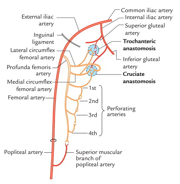
The arterial anastomoses on the back of thigh are as follows:
1. Longitudinal arterial anastomosis: The main arterial supply to the back of the thigh is originated from the perforating branches of the profunda femoris artery. The perforating arteries pierce the adductor magnus and split into ascending and descending branches. Close to the posterior aspect of the insertion of adductor magnus a chain of longitudinal arterial anastomosis is composed by the ascending and descending branches of 4 perforating arteries. The ascending branch of first perforating artery takes part in the formation of cruciate anastomosis and lowest branch anastomose with the superior muscular branch of the popliteal artery.
2. Trochanteric anastomosis: It’s situated in the trochanteric fossa.
The trochanteric anastomosis is composed by these arteries:
- Ascending branch of the medial circumflex femoral artery.
- Ascending branch of lateral circumflex femoral artery.
- Descending branch of the inferior gluteal artery.
- Descending branch of the superior gluteal artery.
3. Cruciate anastomosis: It’s situated in the upper part of the back of femur in the level of the lesser trochanter.
The cruciate anastomosis is composed by the following arteries:
- Transverse branch of the medial circumflex femoral artery.
- Transverse branch of the lateral circumflex femoral artery.
- Ascending branch of the very first perforating artery.
- Descending branch of the inferior gluteal artery.
Clinical Significance
Clinical importance of longitudinal arterial anastomosis on the back of thigh: It gives a collateral channels of blood supply to the lower limb avoiding the external iliac and femoral arteries, example, in ligation of femoral artery above the origin of profunda femoris artery it keeps an efficient blood supply through collateral circulation.

 (48 votes, average: 4.69 out of 5)