Hamstring muscles are situated at the back of the thigh (Latin – “Ham” – back of the thigh). But, still Biceps femoris, which is normally the short headed muscle isn’t included in hamstring muscle group.
The hamstring muscles are:
- Semitendinosus.
- Semimembranosus.
- Biceps femoris (long head).
- Ischial head of adductor magnus.
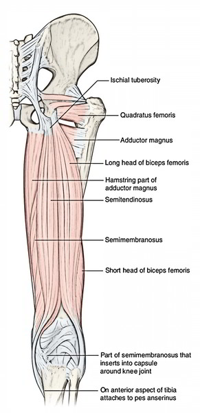
The characteristic features of hamstring muscles are:
- All originate from the ischial tuberosity.
- All are added into 1 of the bones of the leg.
- All are furnished by tibial part of the sciatic nerve.
- All are flexors of the knee and extensors of the hip joint.
Points to be noticed
- The posterior thigh muscles were called “hamstrings” because their tendons on the rear of knee are accustomed to hang up hams (hip and thigh regions of critters viz., pigs.).
- The adductor magnus reaches only up to the adductor tubercle of the femur, but is comprised amongst the hamstrings group of muscles since the tibial collateral ligament of the knee joint morphologically represents the degenerated tendon of the muscle, that is connected below on the tibia.
Origin, Insertion, And Nerve Supply of The Muscles On The Back of thigh
| Muscle | Origin | Insertion | Nerve supply |
|---|---|---|---|
| Biceps femoris (a) Long head | (a) Long head: From lower medial part of upper quadrilateral area of ischial tuberosity | Into the head of the fibula in front of its styloid process | (a) Long head, by the tibial part of the sciatic nerve (L5; S1, S2) |
| (b) Short head | (b) Short head: From lateral lip of the linea aspera and from the upper two-third of the lateral supracondylar line | (b) Short head by the common peroneal part of the sciatic nerve (L5; S1, S2) | |
| Semitendinosus | From the lower medial part of upper quadrilateral area of the ischial tuberosity, | Into the upper part of the medial surface of the tibia | Tibial part of the sciatic nerve (L5; S1, S2) |
| Semimembranosus | From the upper lateral part of upper quadrilateral area of ischial tuberosity | Into the horizontal groove on the posterior aspect of the medial condyle of the tibia | Tibial part of the sciatic nerve (L5; S1, S2) |
| Ischial part of adductor magnus | From inferolateral aspect of the ischial tuberosity | Into the adductor tubercle | Tibial part of the sciatic nerve (L5; S1, S2) |
Biceps Femoris
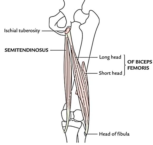
The biceps femoris muscle as its name indicates is composed of 2 heads- long head and short head.
Origin
- Long head, originates from lower medial part of the upper quadrilateral area of the ischial tuberosity in common with the semitendinosus and also from the lower part of the sacrotuberous ligament.
- Short head, originates from the lower part of the lateral lip of the linea aspera and upper 2-third of the lateral supracondylar line.
Insertion
The 2 heads unify in the lower third of the thigh to create a conjoint tendon, which slopes downward and laterally to be added on to the head of fibula in front of the styloid process. Just before insertion the tendon is either folded around or divide by the fibular collateral ligament.
Nerve Supply
- Long head, by the tibial part of the sciatic nerve.
- Short head, by the common peroneal part of the sciatic nerve.
Semitendinosus

It’s thus termed because its lower half is tendinous.
Origin
It originates alongside the long head of biceps femoris from lower medial part of the upper quadrilateral area of the ischial tuberosity. It’s fleshy in the upper part and creates a cord-like tendon in the lower part, which is located on the semimembranosus muscle.
Insertion
In the lower part of the back of thigh, it diverges medially and enters behind the medial condyle of the femur and after that curves downward and forward to be added into the upper part of the medial surfaces of the tibia supporting the insertion of sartorius and gracilis muscles.
Nerve Supply
It’s by tibial part of the sciatic nerve.
Semimembranosus
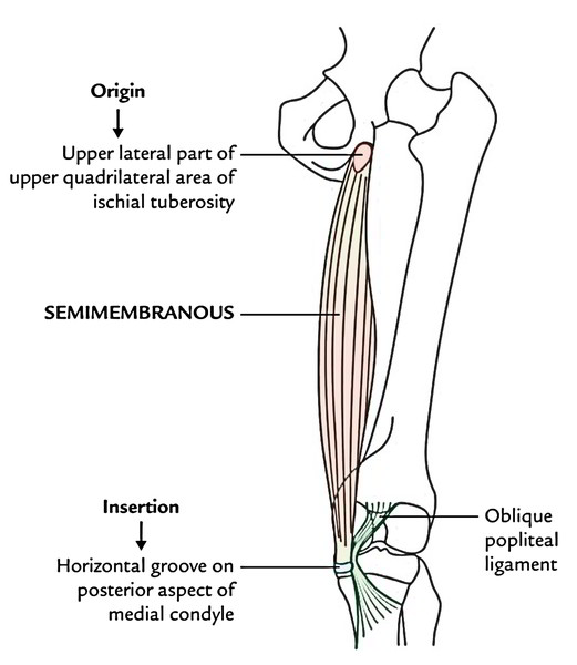
It’s so called because this broad muscle is half membranous.
Origin
It appears from the upper lateral part of the quadrilateral area of the ischial tuberosity. It’s membranous in the upper half and fleshy in the lower half. It is located deep to semitendinosus.
Insertion
The fleshy part converges below to create a tendon, which is added into a horizontal groove on the rear of the medial condyle of tibia.
The tendon of insertion provides rise to the subsequent 3 growths:
- A fibrous band stretches upward and laterally behind the posterior aspect of the capsule of knee joint to create oblique popliteal ligament of the knee.
- A fibrous growth goes downward and laterally through the fascia covering the popliteus to be connected to the soleal line.
- Some fibres descend downward for connection on the medial border of the upper part of tibia behind the tibial collateral ligament.
A synovial bursa lines deep to the tendon of insertion of semimembranosus which might convey with all the synovial cavity of the knee joint.
Nerve Supply
It’s by tibial part of the sciatic nerve.
Ischial Head of The Adductor Magnus
Origin
- It appears from the inferolateral part of the ischial tuberosity.
Insertion
- It descends just about vertically downward to be added on the adductor tubercle.
Nerve Supply
- It’s by tibial part of the sciatic nerve.
Actions of The Hamstring Muscles
- They’re the main flexors of the knee joint and poor extensor of the hip joint; nonetheless, the 2 Actions can not be performed maximally at the same time.
- The hamstrings are the hip extensors during walking on the flat earth when the gluteus maximus exercise minimal task.
- The actions of hamstring muscles limits the range of motion (ROM) of the hip flexion when the knee is extended, viz., during toe touching.
Clinical Significance
Clinical importance of hamstring muscles:
If hamstring muscles are paralyzed, the patient tends to drop forwards as the gluteus maximus muscle can not keep the essential tone to stand erect.
In ancient times, the soldiers utilized to slash the rear of the knees of horses of their rivals to be able to cut the tendons of hamstring muscles, to bring the horse and its rider down. They also utilized to cut the hamstring tendons of soldiers in order that they couldn’t run. This was referred to as “hamstringing” the enemy.

 (59 votes, average: 4.78 out of 5)