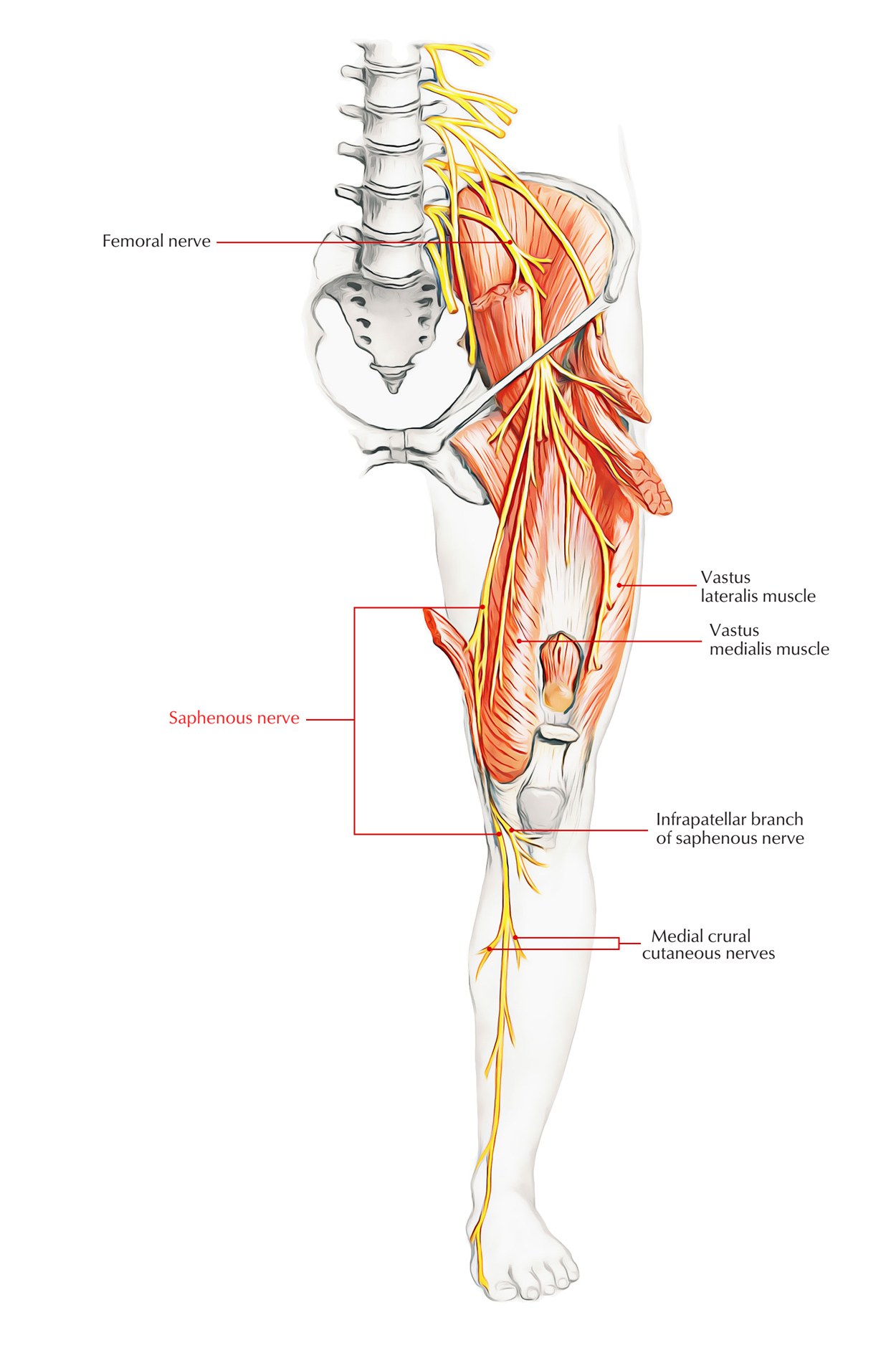
The saphenous nerve is the longest cutaneous division of the femoral nerve and saphenous nerve is the terminal sensory branch of the posterior division of the femoral nerve. The canal, which lies in the center third of the medial thigh, likewise consists of the femoral artery and vein.

Saphenous Nerve
Origin of Saphenous Nerve
Saphenous nerve arises via the canal to become subcutaneous around 10 cm proximal to the medial femoral condyle and originates near the inguinal ligament and comes down inside the quadriceps muscles in the subsartorial (Hunter’s) canal.
Insertion of Saphenous Nerve

Saphenous Nerve: Insertion
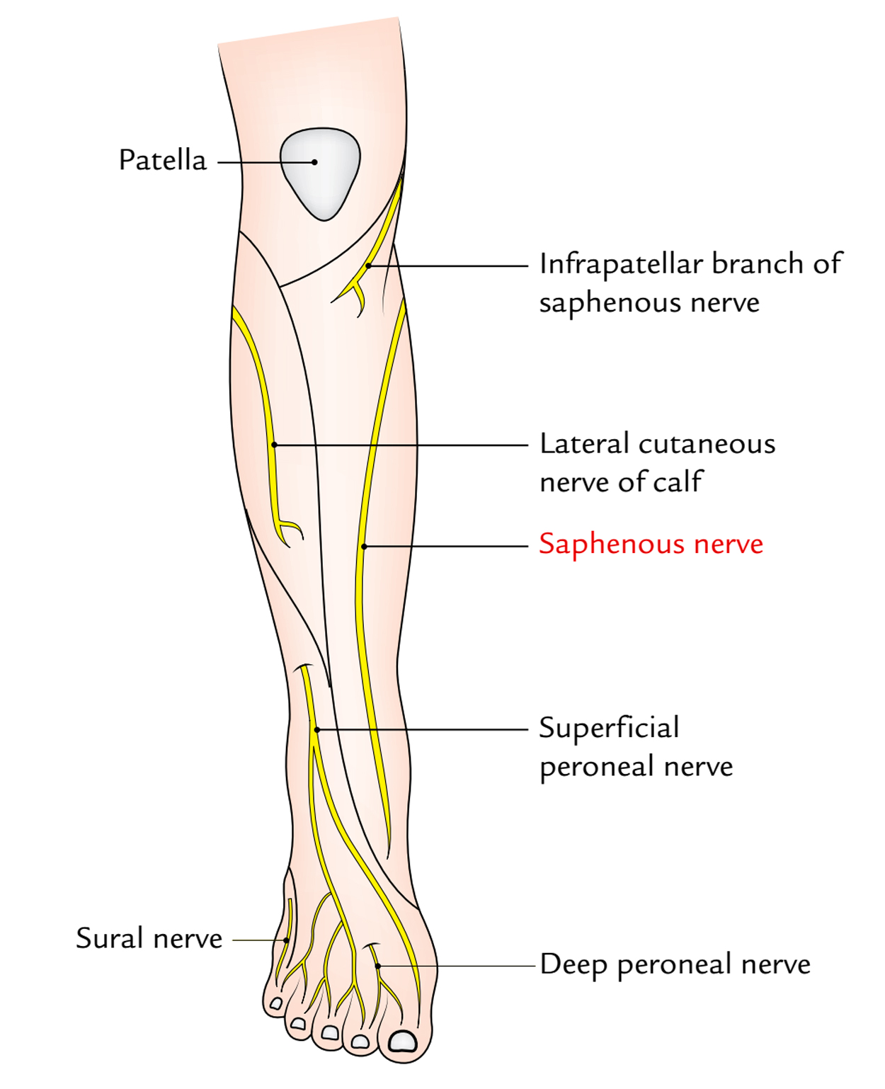
- Saphenous nerve supplies the skin on the medial side of the knee, leg, and medial border of the foot approximately the ball of the big toe.
- Saphenous nerve enters the deep fascia on the medial side of the knee and follows the great saphenous vein, either in front or behind it
- The saphenous nerve goes mainly down the medial aspect of the leg on the medial head of the gastrocnemius, together with the greater saphenous vein.
- Among the larger branches of saphenous nerve, the infrapatellar branch intercrosses medially to laterally throughout the anterior element of the leg near the level of the inferior pole of the patella. This is often transected while anterior approaches of the knee.
Function of Saphenous Nerve
The saphenous nerve supplies cutaneous sensation via the anteromedial element of the knee, medial calf, medial malleolus, and part of the medial arch of the foot, together with its two primary departments, the sartorial and infrapatellar nerves
Clinical Significance of Saphenous Nerve
Entrapment
- Saphenous nerve entrapment can take place as it takes a trip through the subsartorial (I turner’s) canal.
- Compression or thrombosis of the superficial femoral artery inside the adductor canal can trigger claudication pain in the lower leg.
- Neural compression via the femoral vessels and adductor niagnus tendons, that is more proximal to the subsartorial (Hunter’s) canal, has actually likewise been reported.
- Other case reports explain compression of the nerve due to an osteochondroma and via sirring astride and grasping a surf board in between the knees.
Spontaneous Saphenous Neuralgia
Via nerve compromise in the subsartorial canal where the nerve crosses the femoral artery ostensibly and penetrates the roof of the canal spontaneous saphenous neuralgia might take place.
Medial knee and leg pain outcome, and inflammation over the subsartorial canal exists.
Injury while Surgery
- The infrapatellar branch of saphenous nerve might be hurt while arthroscopy or other knee operations, by accidental trauma, or by nerve compression.
- The saphenous nerve might be harmed at the knee after medial arthrotomy or arthroscopy, in association with coronary artery bypass graft surgery, and by lacerations.
- Saphenous nerve is most prone to injury where it pierces the aponeurotic roof of the adductor canal above the knee.
- Surfers who sit astride their boards and grip the boards in between their knees might establish nerve compression.
Meniscal Cyst Compression
A meniscal cyst can compress the nerve at the knee. In all these syndromes, the only indication is a sensory disruption that impacts the medial side of the lower leg.
Gonyalgia Paresthetica
Gonyalgia paresthetica is typically accompanied by acute pain listed below and lateral to the knee and generally establishes insidiously without intense trauma.
Damage to the infrapatellar branch of the saphenous nerve leads to loss of sensation and paresthesias in the skin over the patella, when flexing the knee, occasional pins-and-needles sensations are produced

 (51 votes, average: 4.73 out of 5)