The Ulna is the medial bone of forearm and is homologous to the lateral bone of leg– the fibula. The ulna is attached to by muscles in the arm and forearm to perform movements of wrist, hand and the arm. The end of the ulna presents a large notch-trochlear, or the semilunar, notch – that articulates with the trochlea of the humerus (upper arm bone) to form the elbow joint.
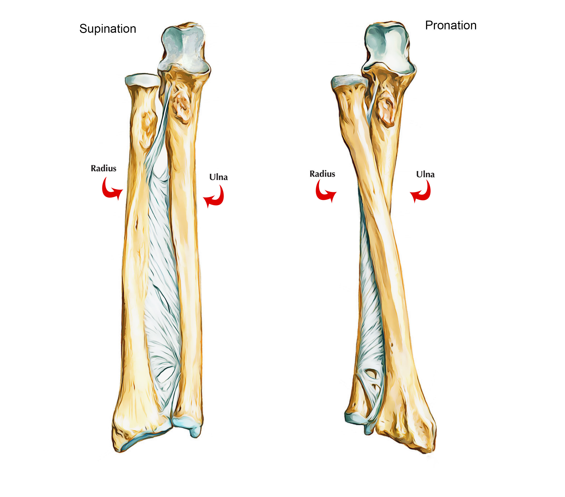
Ulna
Parts of the Ulna Bone
The ulna is a long bone and is composed of three parts : upper end, lower end, and shaft.
Upper End of The Ulna
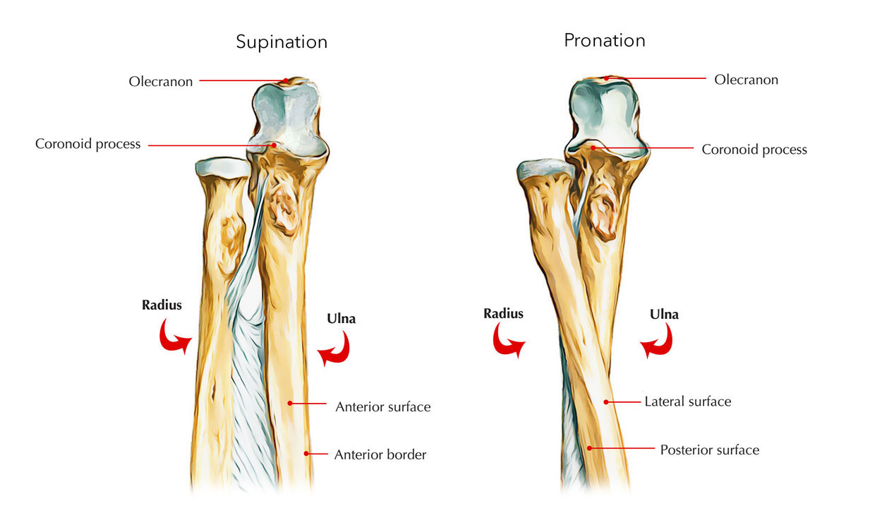
The upper end of the ulna is expanded and hook-like with concavity of hook facing forwards. The concavity of upper end (trochlear notch) is situated between large olecranon process above and the small coronoid process below.
The upper end has 2 processes: coronoid and olecranon, and 2 notches: trochlear and radial.
Ulna – Upper End
Processes
Olecranon process
It projects upwards from the upper end and bends forward at its top like a beak. It has the following 5 surfaces:
Upper surface
- Its rough posterior two-third offers insertion to the triceps brachii.
- Capsular ligament of elbow joint is connected anteriorly near its margins.
- A synovial bursa is located between the tendon of triceps and capsular ligament.
Anterior surface
- It is smooth and forms upper component of the trochlear notch.
Posterior surface
- It makes a subcutaneous triangular area.
- A synovial bursa (subcutaneous olecranon bursa) is located between posterior surface and skin.
- Its upper part offers connections to three structures: (a) ulnar head of flexor carpi ulnaris (origin), (b) posterior, and (c) oblique bands of ulnar collateral ligament.
Coronoid process
It is bracket-like projection coming from the front of the upper end of the ulna, below the olecranon process. It has 4 surfaces: superior, anterior, medial, and lateral.
Superior surface: It is smooth and makes up the lower part of trochlear notch.
Anterior surface: It is triangular in shape.
- Its lower corner presents an ulnar tuberosity.
- Brachialis muscle is inserted to the entire of the anterior surface including ulnar tuberosity.
- The medial margin of the anterior surface is sharp and has a tubercle at its upper end called sublime tubercle. The medial margin offers connection to the following structures starting with proximal to distal:
1. Anterior band of ulnar collateral ligament.
2. Oblique band of ulnar collateral ligament.
3. Humero-ulnar head of flexor digitorum superficialis.
4. Ulnar head of pronator teres.
5. Ulnar head of flexor pollicis longus.
Medial surface: It provides origin to flexor digitorum profundus.
Lateral surface
- The upper part of this surface possesses a radial notch for articulation with the head of the radius.
- The annular ligament is connected to the anterior and posterior margins of the radial notch.
- The lower part of the lateral surface below radial notch has a depressed area referred to as supinator fossa, which includes radial tuberosity in the course of supination and pronation.
- Supinator fossa is bounded behind by supinator crest. Supinator crest and adjoining part of supinator fossa gives origin to the supinator muscle.
Notches (Articular Surfaces)
Trochlear notch
- It is C-shaped (semilunar) and articulates along with the trochlea of humerus.
- It has a non-articular strip at the junction of its olecranon and coronoid parts.
- Its superior, medial, and anterior margins offer connection to capsule of the elbow joint.
Radial notch
It articulates along with the head of radius to form the superior radio-ulnar joint.
Shaft of The Ulna
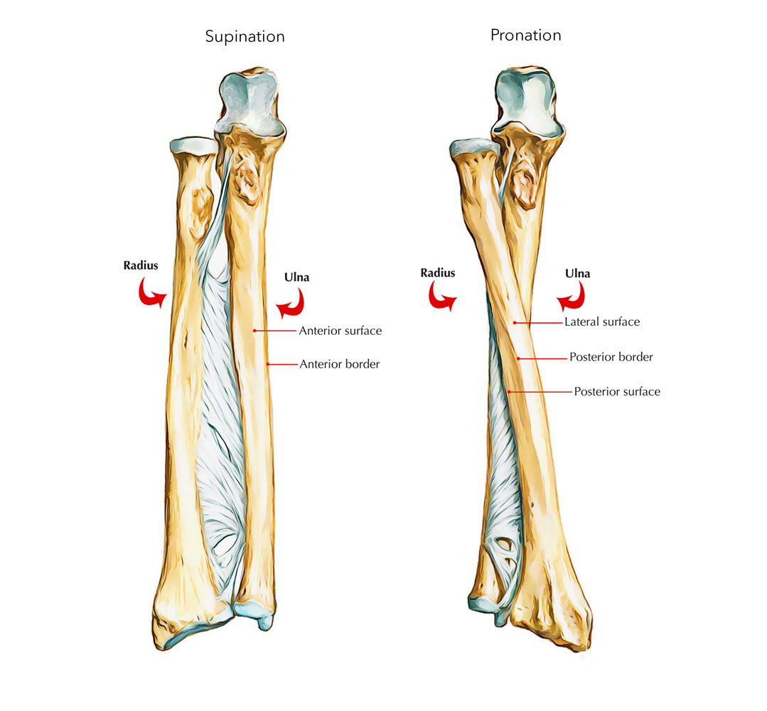
The long shaft of the ulna is between the upper and lower ends. Gradually its thickness decrease from above downwards throughout its length. The lateral border (interosseous border) is sharp crest-like.
Ulna bone’s shaft has three borders (lateral, anterior, and posterior) and three surfaces (anterior, medial, and posterior).
Shaft of Ulna
Borders
Lateral (interosseous) border
- It is sharpest and is continuous above with the supinator crest.
- It is ill-defined below.
- Interosseous membrane is connected to this border with the exception of its upper part.
Anterior border
- It extends from the medial side of the ulnar tuberosity to the base of styloid process.
- It is thick and round.
- It upper three-fourth gives origin to flexor digitorum profundus.
Posterior border
- It begins with the apex of triangular subcutaneous area on the back of olecranon process and descends to the styloid process.
- It is subcutaneous throughout, hence can be palpated along its entire length.
- It provides connection to 3 muscles by a common aponeurosis. The muscles are:
- Flexor digitorum profundus.
- Flexor carpi ulnaris.
- Extensor carpi ulnaris.
Surfaces
Anterior Surface
- It is located between anterior and interosseous borders.
- The flexor digitorum profundus comes up from its upper three-fourth.
- The pronator quadratus arises from an oblique ridge on the lower one-fourth of this particular surface.
- The nutrient foramen is situated a little above the middle of this surface and is directed upwards.
Medial surface
- It is located between the anterior and posterior borders.
- The flexor digitorum profundus arises from the upper two-third of this surface.
Posterior surface
- It is located between posterior and interosseous borders.
- It is divided into smaller upper part and large lower part by an oblique line, which begins at the junction of upper and middle third of posterior border and runs in the direction of the posterior edge of radial notch.
- Area above the oblique line receives insertion of anconeus muscle.
- Area below the oblique line is divided into larger medial and smaller lateral parts by a faint vertical line. The lateral part offers connection to three muscles form proximal to distal as follows:
- Abductor pollicis longus in the upper one-fourth.
- Extensor pollicis longus in the middle one-fourth.
- Extensor indicis in the next one-fourth.
- The distal one-fourth is without any connections.
Lower End of the Ulna
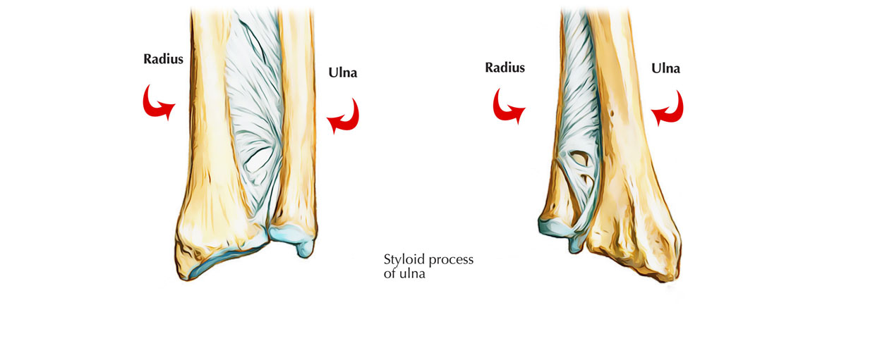
The lower end of the ulna is somewhat expanded and has a head and styloid process. The styloid process is posteromedial to the head.
The ulna appears like a pipe wrench with olecranon process appearing like the upper jaw, the coronoid fossa, the lower jaw, and the trochlear notch the mouth of the wrench.
The lower end of the ulna is composed of head and styloid process.
Ulna – Lower End
Head
- It provides a convex articular surface on its lateral side for articulation with the ulnar notch of radius to make up the inferior radio-ulnar joint.
- Its inferior surface is smooth and separated from wrist joint by an articular disc of inferior radio-ulnar joint.
Styloid Process
- It projects downwards from the posteromedial aspect of the head of ulna.
- Its tip offers connection to medial collateral ligament of wrist joint.
- The apex of triangular articular disc is connected to the depression between head and base of styloid process.
- Tendon of extensor carpi ulnaris is located in the groove between the back of the head of ulna and styloid process.
Anatomical Position And Side Determination
The side of ulna can be identified by keeping the bone vertically in such a manner that:
- The broad hook-like end is directed upwards.
- The sharp crest-like interosseous border of shaft is directed laterally.
- The concavity of the hook-like upper end and the coronoid process are facing forwards.
Ossification of The Ulna Bone
The ulna bone ossifies from the 3 main centres: one primary centre for the shaft and two secondary centres, one each for the lower end and the upper end.
Primary centre
It shows up in the mid-shaft during eighth week of IUL.
Secondary centres
Upper end
- Appearance: 9 years (upper part of trochlear surface and top of olecranon process).
- Fusion: 18 years.
Lower end (middle of head)
- Appearance: 6 years.
- Fusion: 20 years.
Clinical Relevance
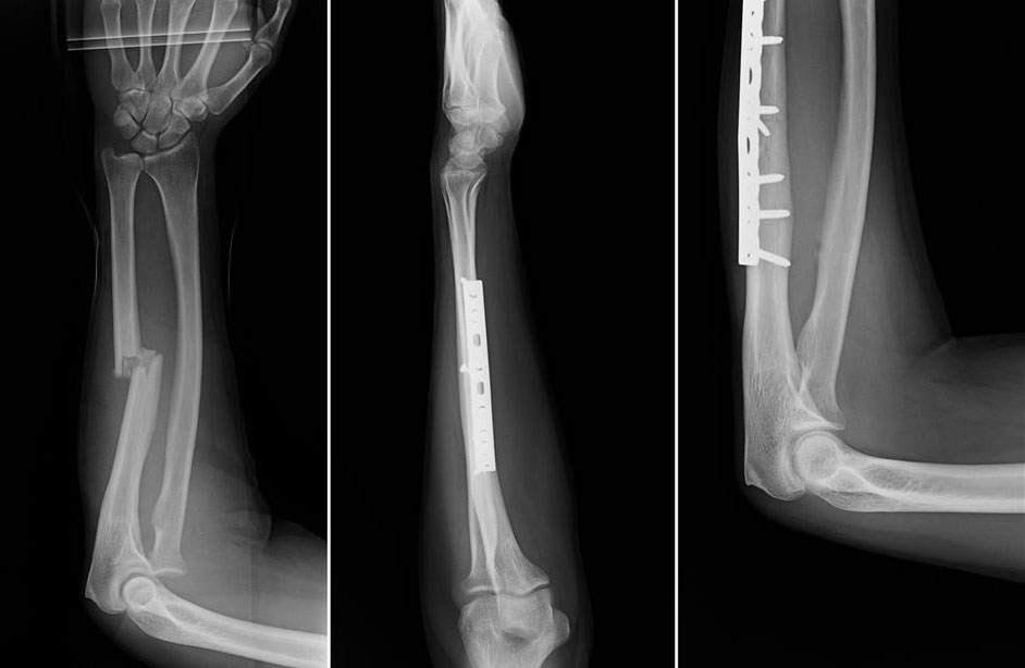
Fractured Ulna
- When the elbow is fully extended, the tip of olecranon process and medial and lateral epicondyles of the humerus hinge on a same horizontal line. When the elbow is fully flexed the three bony points form an equilateral triangle. In dislocation of elbow this relationship is disturbed.
- Ulna stabilizes the forearm by gripping the lower end of humerus by its trochlear notch and offers foundation for radius to produce supination and pronation at superior and inferior radio-ulnar joints.
- The fracture of upper third of shaft of ulna with dislocation of radial head at superior radio-ulnar joint is referred to as Monteggia fracture dislocations.
- The fracture of lower third of the shaft of radius connected with dislocation of inferior radio-ulnar joint is referred to as Galeazzi fracture dislocation.
- A fracture of the shaft of ulna as a result of direct injury when a night watchman reflexly raises his forearm to ward off the blow of the stick is termed night-stick fracture.

 (61 votes, average: 4.84 out of 5)