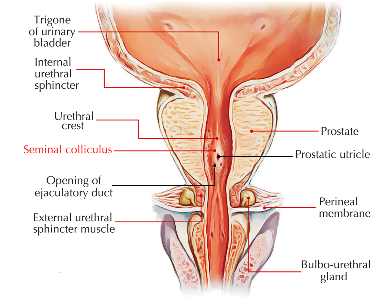
The Verumontanum is an elevated rim of the prostatic urethra on the posterior side. The urethral lining mucosa of the vemmontanum is generally urothelial, however might occasionally with an excessive papillary design be prostatic – type epithelium. Generally the utricle shows an ostium on the side of the vemmontanum. Prostatic ducts from the peripheral and central zones as well as the ejaculatory ducts end on the side of the vemmontanum.
The Verumontanum (also known as seminal colliculus) is a structure situated on the floor of the posterior urethra, which marks the border in between the membranous as well as the prostatic section. On both sides of the urethral ridge, the orifices of the prostatic ducts open, and the ejaculatory ducts’ orifices along with prostatic utricle orifice can be discovered on the upper edge. Generally it has a length of 15-17 mm and a height of 3 mm, while there many variants of both sizes and shape.

Seminal Colliculus
Structure
In men, the seminal colliculus is utilized to identify the location of the prostate gland throughout transurethral transection of the prostate. The structure of the verumontanum consists of striated muscle fibers of the external sphincter, interwoven with smooth muscle tissue from the urethral wall. Marking a crossroads in between the urinary and the genital tracts, verumontana might be impacted in a variety of pathological lesions. Likewise, the spermatic path can be discovered for diagnostic functions via the verumontanum.
Location and Attachments
- On each part of the prostatic utricle is the entrance of the ejaculatory duct of the male reproductive system. A small blind-ended pouch – the prostatic utricle opens up over the center of the seminal colliculus. For that reason the connection in between the urinary and reproductive tracts in men happens in the prostatic part of the urethra.
- The verumontanum is a crucial anatomic landmark for pathology within a congenital anomaly referred to as posterior urethral valves.
- The structure has the tendency to move caudally, or downward, in hypospadia disorders and also is then seen in the round or penile portion of the urethra.
- The prostatic portion of the urethra is 3 to 4 cm long and is encompassed by prostate. In this particular region, the lumen of the urethra is denoted by a longitudinal midline fold of mucosa. The depression at each side of the crest is the prostatic sinus: the ducts of the prostate clear within these two sinuses.
Classification
One category divides verumontana into physiological verumontana (not clinically considerable), verumontana with urinary repercussions, or verumontana considerable for genital pathology.
Physiological verumontana might have different sizes and shapes, a morphological category dividing them into:
- Flat.
- Elongated.
- Humpback.
- Very long.
- Helmet shaped.
- Verumontanum with the element of a diverticulum.
- Other types: represented by combined or intermediate types in between the classifications most often experienced.
The verumontanum can be hypertrophied in some patients, and this large structure might have urinary effect. An element of the spermatic ducts joining verumontanum might recommend a genital pathology.
Clinical Significance
The endoscopic method of this structure, for both indicative and healing intent, might turn out vital in the management of male infertility. Throughout endoscopic surgery, it acts as a landmark of the striated sphincter and, unconditionally, of the lower limitation of the intervention.

 (46 votes, average: 4.85 out of 5)