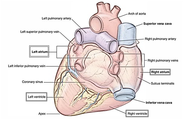
The heart is composed of 4 chambers, viz.
A. Right atrium.
B. Right ventricle.
C. Left atrium.
D. Left ventricle.
The 2 atrial chambers are divided from every other by a vertical septum the interatrial septum and the 2 ventricular chambers are divided from every other by a vertical septum the interventricular septum.
The right atrium interacts with the right ventricle via right atrioventricular orifice, that is guarded by 3 cusps.
The left atrium interacts with all the left ventricle via the left atrioventricular orifice, that is guarded by 2 cusps.
The walls of the chambers of the heart are composed of cardiac muscle the myocardium, that is covered externally by the serous membrane- the epicardium and lined internally by endothelium- the endocardium.
The atria are thin walled as compared to the ventricles and have little contractile power.
Demarcation Of Chambers Of The Heart On The Surface
On the surface the chambers of the heart are demarcated or delineated by the following 3 grooves:
A. Coronary sulcus (atrioventricular groove).
B. Anterior interventricular sulcus.
C. Posterior interventricular sulcus.
Coronary Sulcus (Atrioventricular Groove)
It encircles the heart and separates the atria from the ventricles. It’s deficient anteriorly because of the root of pulmonary trunk.
The atrioventricular groove is split into anterior and posterior parts.
The Anterior Part Is Composed Of Left And Right Halves
The right half of the anterior part runs downwards and to the right between the right atrium and right ventricle and lodges right coronary artery.
The left anterior part of AV groove intervenes between the left auricle and left ventricle. It lodges circumflex branch of left coronary artery.
The posterior part of AV groove intervenes between the base and the diaphragmatic surface of the heart. It lodges coronary sinus.
Anterior And Posterior Interventricular Sulci
They separate the left and right ventricles. The anterior interventricular sulcus is on the sternocostal surface of the heart and lodges anterior interventricular artery and great cardiac vein. The posterior interventricular groove is on the diaphragmatic surface and lodges posterior interventricular artery and middle cardiac vein.
The meeting point of interatrial groove, posterior interventricular groove, and posterior part of atrioventricular groove is referred to as crux of the heart.
Circulation Of Blood
Functionally, the heart is created from 2 muscular pumps – the left and right. The right pump is composed of right atrium and right ventricle while the left pump is composed of left atrium and left ventricle. The right pump is responsible for pulmonary circulation and the left pump is responsible for systemic circulation as follows:
1. The right atrium gets deoxygenated blood from the whole body via superior and inferior venae cavae. The blood flows from right atrium into right ventricle via right atrioventricular orifice. The blood is prevented from regurgitating back to the atrium by means of right atrioventricular valve. The right ventricle contracts and propels the blood into the pulmonary trunk, pulmonary arteries, and finally into the lung where blood is oxygenated (pulmonary circulation).
2. The left atrium gets the oxygenated blood from lungs via 4 pulmonary veins. The blood from left atrium flows into left ventricle via left atrioventricular orifice. The blood is prevented from regurgitating back to the atrium by means of left atrioventricular valve. The left ventricle strongly contracts and propels the blood into the ascending aorta and after that into the systemic circulation.
3. The right ventricle is required to pump the blood via a relatively low-resistance vascular bed, on the other hand the left ventricle is required to pump the blood via a relatively high resistance peripheral vascular bed.
4. The muscular wall of the left ventricle is, for that reason, much thicker than that of the right ventricle.
Right Atrium
The right atrium is somewhat quadrilateral chamber situated behind and to the right side of the right ventricle. It is composed of a main cavity and a small outpouching named auricle.
External Features
A. The right atrium is elongated vertically and gets superior vena cava (SVC) at its upper end and the inferior vena cava (IVC) at its lower end.
B. The upper anterior part is prolonged to the left to create the right auricular appendage, the right auricle. The margins of the auricle are notched. The right auricle overlaps the roots of the ascending aorta completely and infundibulum of the right ventricle partly.
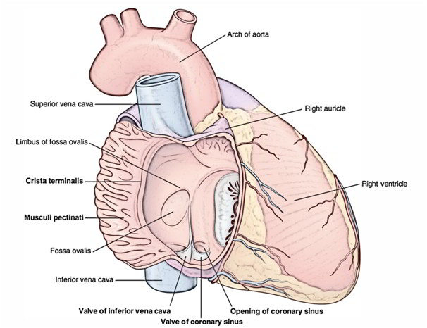
C. A shallow vertical groove referred to as sulcus terminalis extends along the right border between the superior and inferior vena cavae. The upper part of the sulcus includes the sinuatrial (SA) node. Internally it corresponds to crista terminalis.
D. The vertical right atrioventricular groove lodges the right coronary artery and the small cardiac vein.
Internal Features
The interior of the right atrium is split into 2 parts:
A. Main smooth posterior part – the sinus venarum, and.
B. Rough anterior part – the atrium proper. The 2 parts are divided from every other by crista terminalis. The interior of right atrium also presents septal wall of the right atrium.
Septal wall of the right atrium
Developmentally it’s originated from septum primum and septum secundum. The septal wall when viewed from inside the right atrium presents the following features:
A. Fossa ovalis, a shallow oval/saucer-shaped depression in the lower part, created by septum primum. It represents the site of foramen ovale in the foetus.
B. Annulus ovalis/limbus fossa ovalis, creates the distinct upper and lateral margin of the fossa ovalis. It represents the free edge of the septum secundum. Inferiorly the annulus ovalis is continuous with the left end of the valve of IVC.
C. Triangle of Koch, a triangular area bounded in front by the base of septal leaflet of tricuspid valve, behind by anterior margin of the opening of coronary sinus and above by the tendon of Todaro- a subendocardial ridge. The atrioventricular node is located in this triangle.
D. Torus aorticus, an elevation in the anterosuperior part of the septum produced because of bulging of the right posterior (non-coronary) sinus of ascending aorta.
Differences Between The Smooth And Rough Parts Of The Right Atrium
Smooth part (sinus venarum) | Rough part (atrium proper) |
|---|---|
| Developmentally it is derived from right horn of the sinus from primitive atrium venosus | |
| All the venous channels except anterior cardiac veins open into this part (e.g., SVC, IVC, coronary sinus, and venae cordaeminimi) | Presents series of transverse ridges, the musculipectinati, which arise from the crista terminalis and run forwards towards the auricle. The interior of auricle presents reticular sponge-like network of the muscular ridges |
Clinical Significance
The sponge-like interior of right auricle prevents the free blood flow and thus favors the formation of thrombus. The thrombi may dislodge during auricular fibrillation and can cause pulmonary embolism.
Opening Into The Right Atrium
There are number of openings in the right atrium. These regions follows:
A. Opening of SVC: The SVC opens at the upper end of the right atrium and has no valve. It returns the blood to the heart from the upper half of the body.
B. Opening of IVC: The IVC opens at the lower end of the right atrium near the interatrial septum. It’s guarded by a rudimentary non-functioning semilunar valve referred to as valve of the inferior vena cava/Eustachian valve.
C. Opening of coronary sinus: The coronary sinus, which drains the majority of the blood from the heart, opens into the right atrium between the openings of IVC and right atrioventricular orifice. It’s also guarded by a rudimentary non-functioning valve, Thebesian valve.
D. Right atrioventricular orifice (largest opening): It interacts the right atrial chamber with all the right ventricular chamber. It is located anterior to the opening of IVC and is guarded by the tricuspid valve.
E. Many small orifices of small veins: These are the opening of venae cordisminimae (Thebesian veins) and anterior cardiac veins.
During embryonic life, the Eustachian valve guides the blood of IVC to the left atrium via foramen ovale. The IVC returns the blood to the heart from the lower half of the body. A very small projection termed intervenous tubercle (of Lower) is scarcely visible on the posterior wall of the right atrium just below the opening of SVC. During embryonic life it directs the blood of SVC to the right ventricle.
Right Ventricle
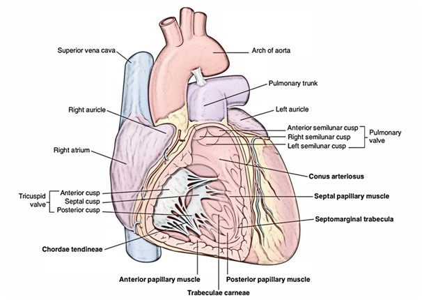
The right ventricle is the thick-walled triangular chamber of the heart which interacts with the right atrium via right atrioventricular orifice and with the pulmonary trunk via pulmonary orifice.
External Features
A. It creates the majority of sternocostal surface and small part of the diaphragmatic surface of the heart. It also creates the inferior border.
B. It is divided from the right atrium by a more or less vertical anterior part of the coronary sulcus/ atrioventricular groove.
Differences Of Inflowing And Outflowing Parts Of The Right Ventricle
| Inflowing lower part | Outflowing upper part |
| It develops from primitive ventricle | It develops from bulbuscordis |
| It is large in size and lies below the supraventricular crest | It is small in size and lies above the supraventricular crest |
| It is rough due to presence of It is smooth and forms upper 1 the muscular ridges—the inch conical part of the right trabeculae carneae. It forms ventricular chamber—the most of the right ventricular infundibulum, which gives rise | |
Internal Features
A. The interior of right ventricle is composed of 2 parts: (a) a large, lower rough inflowing part, and (b) a small upper outflowing part, the infundibulum. The 2 parts are divided from every other by a muscular ridge, the supraventricular crest (infundibuloventricular crest).
B. The cavity of right ventricle is flattened by the forward bulge of the interventricular septum. In transverse section it’s crescent shaped.
C. The wall of the right ventricle is thinner than that of the left ventricle (ratio 1:3).
Trabeculae Carneae Of Right Ventricular Chamber
These are muscular projections which give the ventricular chamber a sponge-like appearance.
Types Of Trabeculae Carneae
Trabeculae carneae are of 3 types: (a) ridges (fixed elevations), (b) bridges (only ends are fixed, the central part is free), and (c) pillars (base is fixed to ventricular wall and apex is free).
Papillary Muscles
These represent the pillars of trabeculae carneae. The papillary muscles project inwards. Their bases are connected to the ventricular wall and their apices are connected by thread-like fibrous cords (the chordae tendinae) to the cusps of the tricuspid valve.
There are 3 papillary muscles in the right ventricle: (a) anterior, (b) posterior (inferior), and (c) septal. The anterior is largest, posterior is small, and septal is generally split into 2 or 3 nipples. The papillary muscles of right ventricle are connected to the cusps of the tricuspid valve.
Moderator Band (Septomarginaltrabeculum)
It’s thick muscular ridge extending from ventricular septum to the base of the anterior papillary muscle, across the ventricular cavity. It conveys the right branch of the atrioventricular bundle (bundle of His), a part of conducting system of the heart. It prevents the over distension of right ventricle.
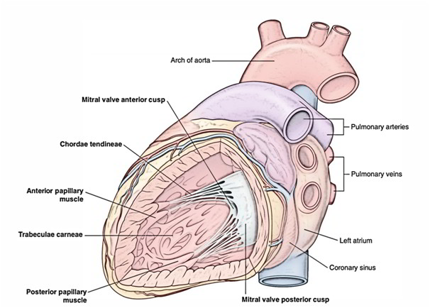
Left Atrium
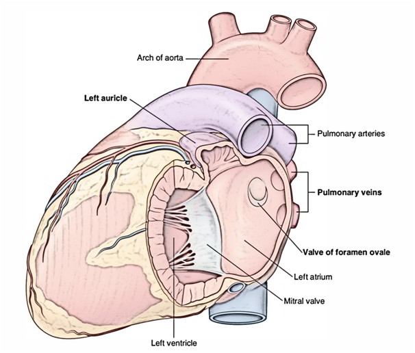
External Features
A. It’s a thin-walled quadrangular chamber situated posteriorly behind and to the left side of right atrium. It creates greater part (left 2/3rd) of the base of the heart.
B. Its upper end is prolonged anteriorly to create the left auricle, which overlaps the infundibulum of right ventricle.
C. Behind the left atrium is located: (a) oblique sinus of serous pericardium and (b) fibrous pericardium, which separates it from the esophagus.
Internal Features
A. The interior of left atrium is smooth, but the left auricle possesses muscular ridges in the form of reticulum.
B. The anterior wall of left atrial cavity presents fossa lunata, which corresponds to the fossa ovalis of the right atrium.
Openings In The Left Atrium
Openings in the left atrium are as follows:
A. Openings of 4 pulmonary veins in its posterior wall, 2 on every side. They’ve no valves.
B. Number of small openings of venae cordisminimae.
C. Left atrioventricular orifice. It’s guarded by the mitral valve.
Left Ventricle
The left ventricle is thick-walled triangular chamber of the heart which interacts with all the left atrium via left atrioventricular orifice and with the ascending aorta via aortic orifice. The walls of left ventricle are 3 times thicker than that of the right ventricle.
External Features
The left ventricle creates the (a) apex of the heart, (b) small part of the sternocostal surface, (c) majority of the (left 2/3rd) diaphragmatic surface, and (d) the majority of the left border of the heart.
Internal Features
The interior of the left ventricle is split into 2 parts: (a) a large lower rough inflowing part, and (b) a small upper smooth outflowing part the aortic vestibule.
The cavity of the left ventricle is circular in cross section since the interventricular septum bulges into the right ventricle.
Differences Between The Inflowing And OutFlowing Parts Of The Left Ventricle
Inflowing part | Outflowing part |
|---|---|
| It develops from primitive ventricle | It develops from bulbuscordis |
| It lies below the aortic vestibule | It lies between the membranous part of the interventricular septum and anterior cusp of the mitral valve |
| It is rough due to presence It is smooth and forms smooth of trabeculae carneae and small upper part—the aortic forms most of the left vestibule, which gives rise to the ventricular chamber ascending aorta | |
Trabeculae Carneae Of Left Ventricle
The trabeculae carneae of the left ventricle are quite similar to those of the right ventricle but are well developed and present 2 large papillary muscles (anterior and posterior) and no moderator band. The papillary muscles of the left ventricle are connected to the cusps of the mitral valve by chordae tendinae.
Openings In The Left Ventricle
The openings in the left ventricle are as follows:
A. Left atrioventricular orifice.
B. Aortic orifice.
Differences Between The Left And Right Ventricles
Right ventricle | Left ventricle |
|---|---|
| Receives deoxygenated blood from right atrium and pumps it to the lungs through pulmonary trunk | Receives oxygenated blood from left atrium and pumps it to the whole body through aorta |
| Wall of right ventricle is thinner than that of left ventricle (ratio 1:3) | Wall of left ventricle is thicker than that of right ventricle (ratio 3:1) |
| Possesses three papillary muscles (anterior, posterior, and septal) | Possesses two papillary muscles (anterior and posterior) |
| Moderator band present | Moderator band absent |
| Cavity of right ventricle is crescentic in shape in cross section | Cavity of left ventricle is circular in shape in cross section |

 (55 votes, average: 4.55 out of 5)