The ileum is primarily in the right lower quarter and comprises the distal three-filths of the small intestine. Compared to the jejunum, the ileum has thinner walls, few and less prominent mucosal folds (plicae circulares) shorter vasa recta, more mesenteric fat, and more arterial arcades.
Structure
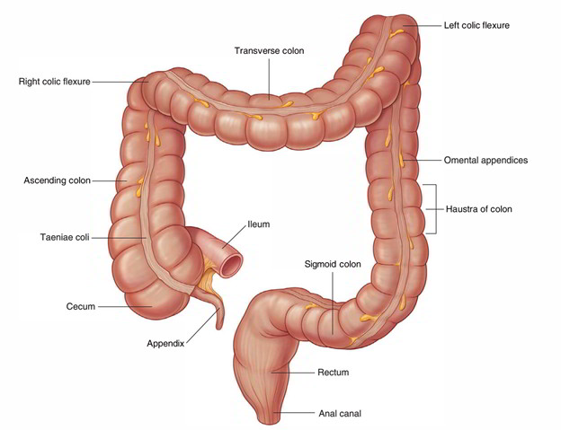
- Where the cecum and ascending colon join together, the ileum opens inside the large intestine.
- Two flaps projecting into the lumen of the large intestine called the ileocecal fold surround the opening.
- The flaps of the ileocecal fold come together at their end creating ridges.
- Musculature via the ileum continues into each flap, forming a sphincter.
- Possible functions of the ileocecal fold consist of avoiding reflux via the cecum towards the ileum, and managing the movement of components via the ileum to the cecum.
Intestinal Glands (Crypts of Lieberkuhn)
They produce digestive enzymes and mucous, between the bases of villi, the epithelium is invaginated in the lamina propria to form intestinal glands.
Lymphatic Follicles
The lamina propria of the mucous membrane consists of two types of lymphatic follicles.
- Solitary lymph follicles: These are dispersed throughout the length of the small intestine and are 1-2 mm in diameter.
- Aggregated lymph follicles: They create circular or oval patches called Peyer’s spots. Each Peyer’s spot length differs from 2 to 10 cm and it includes about 260 singular lymph follicles. They are found alongside around the antimesenteric border. In the distal part of the ileum where Peyer’s spots are much larger and most numerous might expand in the submucosa afterwards breaking the muscularis mucosa. Peyer’s spots are large, oval, and many in the ileum especially in its distal part and small, circular, and fewer in the distal part of the jejunum.
Mesentery of the Small Intestine
It is a broad fan-shaped fold of the peritoneum, which suspends the small intestine (jejunum and ileum) via the posterior abdominal wall. It has the root (connected margin) as well as free margin (intestinal margin).
The duodenojejunal flexure is located to the left side of second lumbar vertebra, whereas ileocaecal junction is located at the right sacroiliac joint. The root is connected to an oblique line throughout the posterior abdominal wall expanding via the duodenojejunal flexure to the ileocaecal junction.
Arterial Supply
The arterial supply to the ileum:
- ileal arteries via the superior mesenteric artery.
- ileal branch via the ileocolic artery (via the superior mesenteric artery).
Venous Drainage
The veins correspond to the branches of superior mesenteric artery and drain within the portal vein, which brings the products of protein and carbohydrates to the liver.
Lymphatic Drainage
The lymph vessels via the small intestine go through a great deal of mesenteric nodes (lymph nodes found in the mesentery) and lastly drain into superior mesenteric nodes found near the origin of superior mesenteric artery.
Nerve Supply
The sympathetic supply is originated from T10-T11 spinal sections via splanchnic nerves and superior mesenteric plexus. The small intestine is innervated by both sympathetic and parasympathetic nerve fibers.


 (121 votes, average: 2.74 out of 5)