There are numerous joints in the foot created between tarsal, metatarsal, and phalangeal bones. The joints of the foot contain intertarsal, tarsometatarsal, intermetatarsal, metatarsophalangeal, and interphalangeal joints. Those joints that are of more functional worth are discussed.
Intertarsal Joints
They’re of 2 types: major and small. Only major intertarsal joints are discussed in the subsequent text.
Subtalar (Talocalcanean) Joints.
There are just two joints between the talus and calcanean: posterior talocalcanean joint and anterior talocalcaneonavicular joint. The posterior talocalcanean joint is frequently designated as subtalar joint. It’s a plane type of synovial joint. It’s created between the concave facet on the inferior outermost layer of the body of talus and convex facet on the middle 1-third of the superior outermost layer of the calcaneum.
Ligaments
These are: (a) fibrous capsule, (b) medial and lateral talocalcanean ligaments, (c) interosseous talocalcanean ligament, and (d) cervical ligament.
The interosseous talocalcanean ligament is thick and extremely powerful. It creates the main bond of union between the talus and calcaneum. It inhabits sinus tarsi and splits the talocalcanean joint from the talocalcaneonavicular joint. It goes obliquely from the sulcus tali to the sulcus calcanei. It becomes tight in eversion. The cervical ligament is lateral to sinus tarsi. It goes upward and medially from upper surface of the calcaneum to the tubercle on the inferolateral aspect of the neck of talus. It becomes tight in inversion.
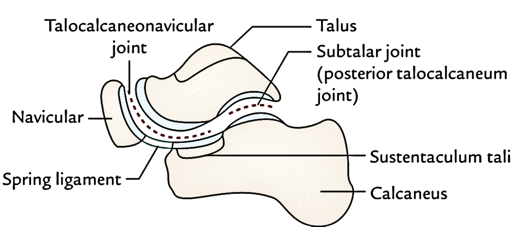
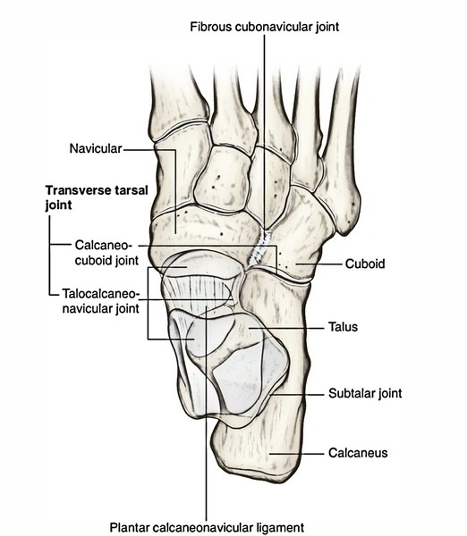
Talocalcaneonavicular Joint
It’s a mixture articulation composed of anterior talocalcanean and talonavicular joints. It’s about a sphere and socket type of synovial joint. The articular surface on the rounded head of the talus fits into the socket created by the calcaneum, navicular, and spring ligament.
Ligaments
These are: (a) fibrous capsule, (b) spring ligament, and (c) medial limb (calcaneonavicular part) of bifurcate ligament.
The spring ligament (plantar calcaneonavicular ligament) is a strong fibrocartilaginous band, which goes from the anterior margin of the sustentaculum tali to the plantar surface of navicular bone between its tuberosity and articular margin. It takes part in creating the socket for the head of the talus. It’s the most essential ligament to keep the medial longitudinal arch. Its upper surface has a triangular fibrocartilagenous facet for the head of the talus. Its plantar surface is supported by the tendon of tibialis posterior medially, and by the tendons of flexor hallucis longus and flexor digitorum longus laterally.
The talocalcaneonavicular joint allows the movements of inversion and eversion.
Calcaneocuboid Joint
It’s a saddle type of synovial joint. The opposed articular surfaces of the calcaneum and cuboid are reciprocally concavoconvex.
Ligaments
The ligaments of the joint are: (a) fibrous capsule, (b) lateral limb (calcaneocuboid part) of bifurcate ligament, (c) long plantar ligament, and (d) short plantar ligament.
The bifurcate ligament is Y shaped. Its stalk is connected to the anterolateral part of the sulcus calcanei. Its medial limb (calcaneonavicular part) is connected to the dorsolateral surface of the navicular bone and its lateral limb (calcaneocuboid part) to the dorsomedial surface of the cuboid bone. The 2 limbs supply support to the joints.
The long planar ligament is the longest ligament. It’s powerful and its relevance in keeping the arches of foot is surpassed only by the spring ligament. It goes from triangular plantar surface of the calcaneum to the lips of the groove on cuboid and beyond it to the bases of the middle 3 metatarsals (2nd to fourth). It converts the groove on theplantar surface of cuboid into a tunnel for the passage of tendon of peroneus longus. Morphologically, it composes the divorced tendon of the gastrocnemius.
The short plantar ligament (plantar calcaneocuboid ligament) is located deep to long plantar ligament. It’s broad and extends from the anterior tubercle of calcaneum to the plantar surface of the cuboid behind its ridge.
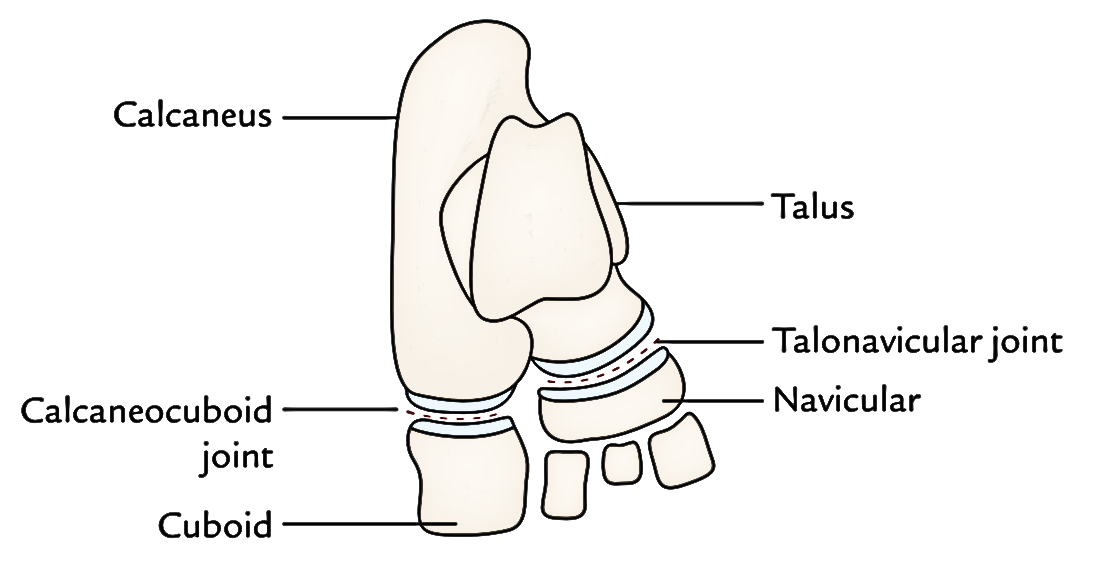
Transverse Tarsal (Midtarsal) Joint
It’s a compound joint being composed of calcaneocuboid and talonavicular joints. Talonavicular is also a part of talocalcaneonavicular joint. The calcaneocuboid and talonavicular joints are grouped together because both are set almost in exactly the same transverse plane. On the other hand, both joints have distinct axes of movements. The movements of midtarsal joint help in inversion and eversion of the foot.
Inversion And Eversion of The Foot
The inversion and eversion and rotational movements of the foot on the talus.
The inversion is a movement where the medial border of the foot is lifted so the sole faces medially.
The eversion is a movement where the lateral border of the foot is lifted so the sole faces laterally.
Joints Taking Part
- Subtalar joint.
- Talocalcaneonavicular joint.
- Transverse tarsal/midtarsal joint- Accessory joint.
Axis of Movements
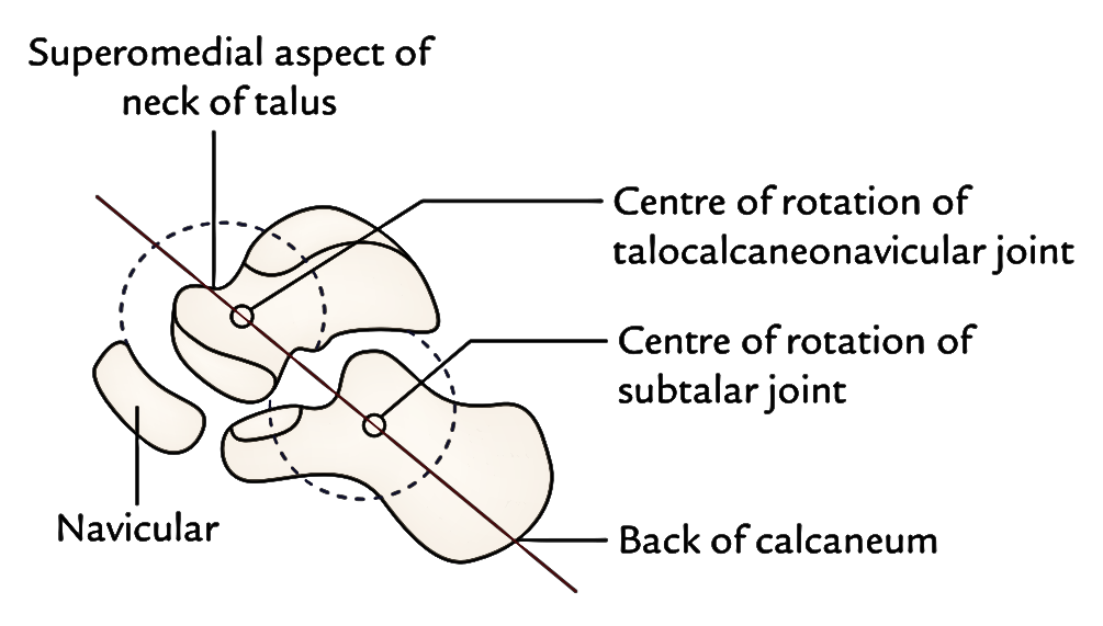
The movements of inversion and eversion take place around an oblique axis which runs forward, upward, and medially going from the rear of calcaneum via the sinus tarsi to appear at the superomedial aspect of the neck of talus.
Range of Movements (Rom)
- The range of movement of inversion is far more than that of eversion (inversion = 30 °, eversion = 20 °)
- The range of these movements is appreciably raised in plantar flexion of the foot because in this position, the narrow posterior part of the trochlear surface of talus takes up the tibiofibular socket (mortise), which allows some level of side to side movement of the talus.
Movements of Inversion and Eversion When the Foot Is off The Earth and When the Foot Is on the Earth
| Movement | Inversion | Eversion |
|---|---|---|
| When foot is off the ground | • Range of motion is more. • Inversion consists of adduction of the forefoot, lateral rotation (supination) of the forefoot, and plantar flexion of the ankle. | • Range of motion is more. • Eversion consists of abduction of the forefoot, medial rotation (pronation) of the forefoot, and dorsiflexion of the ankle |
| When foot is on the ground | • Range of motion is less. • Inversion consists of only lateral rotation (supination) of the forefoot. • Heads of the medial 2 metatarsals are raised | • Range of motion is less. • Eversion consists of only medial rotation (pronation) of the forefoot. • Heads of the lateral 3 metatarsals are raised. |
Movements of Inversion And Eversion
Muscles | ||
|---|---|---|
| Movements | Principal muscles | Accessory muscles |
| Inversion (ROM = 30°) | 1.Tibialis anterior. | 1.Flexor hallucis longus. |
| 2.Tibialis posterior. | 2.Flexor digitorum longus | |
| Eversion (ROM = 20°) | 1.Peroneus longus. 2.Peroneus brevis. | Peroneus tertius |
Muscles Creating Movements
The movements of inversion and eversion and muscles creating them are provided in Table 32.6.
Functional Importance
The movements of inversion and eversion are essential for walking on uneven and sloping grounds. They significantly help the foot in correcting it to such reasons. When feet are supporting the body weight, these movements take place in a modified create termed supination and pronation.
Tarsometatarsal Joints
All these are plane type of synovial joints. They may be joined by dorsal, plantar, and interosseous tarsometatarsal ligaments. They’re 5 in number. The very first joint possesses another cavity, the 2nd and third together have 1 cavity. These joints allow only small gliding movements.
Metatarsophalangeal Joints
All these are ellipsoidal type of the synovial joints. They’re joined by the capsular, collateral, plantar, and deep transverse metatarsal ligaments. 2 collateral ligaments reinforce the sides of every joint. These joints allow the small gliding movements. The deep transverse ligaments (4 in number) attach the plantar ligaments of adjacent metatarsophalangeal joints.
Movements
These joints allow dorsiflexion, plantar flexion, adduction, and abduction.
- The range of dorsiflexion is more (50-60 °)than that of plantar flexion (30-40 °).
- The axis of adduction and abduction of the toes goes through the least mobile 2nd metatarsal bone. (cf. The axis of adduction and abduction of fingers goes through the third metacarpal bone.).
Interphalangeal Joints
All these are typical hinge joints. They’re linked by the capsular and collateral ligaments.
Movements
These joints allow dorsiflexion and plantar flexion of distal phalanges.
Test Your Knwoledge
Joints of Foot

 (57 votes, average: 4.67 out of 5)