The human foot is often called a wonder of nature’s architecture. it is one of the greatest examples of structural adaptation towards change in environment and conditions. Human foot has undergone a lot of phenotype change due to external and internal changes of conditions
The foot performs 2 major functions:
- It functions as a stage to support the body weight during standing position.
- It functions as a lever to propel the body forward during walking, running, and leaping.
To carry through the very first function, the foot was made in the creation of elastic arches. These arches are segmented so they can endure the strain of weight and pushes at the optimum level.
To carry through the next function, the foot is so built that it’s transformable in a lever. The segmented arched lever converts the foot into a spring, that is ideally suited for its functions.
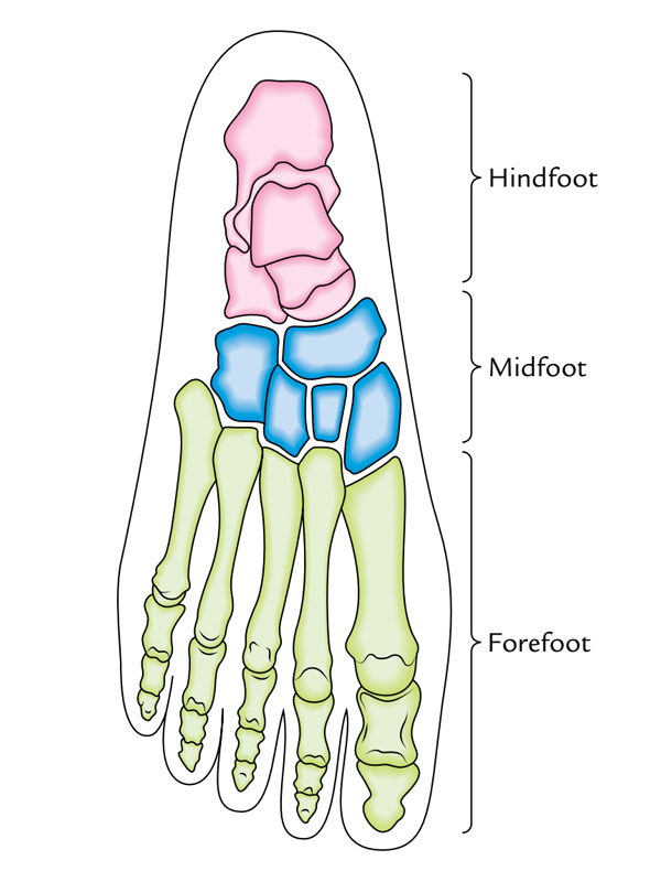
The foot and its bones are split into the following 3 anatomical and functional sections:
- The hindfoot includes talus and calcaneus.
- The midfoot includes navicular, cuboid, and cuneiforms.
- The forefoot includes metatarsals and phalanges.
The skeleton of the foot is arched, both longitudinally and transversely, together with the concavity steered in the direction of the plantar surface (i.e., the bones of the foot are ordered to create the transverse and longitudinal arches). The presence of arches makes the sole concave both anteroposteriorly and transversely. This is best represented in the footprint revealing the weight bearing points of the sole.
During the standing position, the weight of the body is spread among 3 points:
- posteroinferiortuberosity of the calcaneum (heel).
- head of first metatarsal.
- head of fifth metatarsal.
The arches of the foot are present right from beginning, but as a result of presence of excessive subcutaneous fat in the soles, they’re not clear (i.e., hidden) during infancy and youth.
An arched foot is a distinctive feature of guy, which differentiates him from other primates.
Variables Keeping the Arches of the Foot
The people can easily understand the variables keeping the arches of the foot by using the engineering apparatus utilized to support a stone bridge. The apparatus utilized to support a stone bridge are:
- Shape of stones.
- Intersegmental ties (staples).
- Slings.
- Tie beams.
Types of Arches
There are 2 types of arches of the foot- longitudinal and transverse.
- There are 2 longitudinal arches in every foot: (a) medial and (b) lateral.
- There are a series of transverse arches in every foot. At the heads of metatarsals, the transverse arch is whole but posteriorly it creates a half dome, that is completed by its counterpart in the opposite foot. So, in the posterior part of the foot the transverse arch becomes complete when the both feet are held close to every other.

Arches of the Foot: Types of Arches
Longitudinal Arches
Every longitudinal arch has: (a) 2 Pillars, (b) a summit, and (c) joints.

Arches of Foot: Longitudinal Arches
Medial Longitudinal Arch
The medial longitudinal arch is composed by the calcaneum, talus, navicular, 3 cuneiforms, and medial 3 metatarsals.

Arches of Foot: Medial Longitudinal Arch
Pillars
- The medial half of the calcaneum creates the posterior column of the medial longitudinal arch.
- The heads of the medial 3 metatarsals create the anterior column of the medial longitudinal arch.
Summit
The talus is located at the peak of the arch. For that reason, the talus is the keystone of the arch.
Joints
The key joints of the medial longitudinal arch are talocalcaneonavicular and subtalar joints.
Lateral Longitudinal Arch
The lateral longitudinal arch is composed by the calcaneum cuboid and lateral 2 metatarsals. It’s characteristically low and just about touches the earth. It’s involved in receiving and supporting the body weight during walking and running.

Arches of Foot: Lateral Longitudinal Arch
Pillars
The posterior column of the lateral longitudinal arch is composed by the lateral tubercle of the calcaneum and the anterior column is composed by the heads of the lateral 2 metatarsals.
Summit
The peak of the lateral longitudinal arch is located at the level of articular facets on the superior outermost layer of the calcaneum (i.e., at the level of subtalar joint).
Joints
The primary joint of the lateral longitudinal arch is calcaneocuboid joint.
The lateral longitudinal arch being lower and not as mobile in relation to the medial longitudinal arch is accommodated for transmission of weight and pushes.
Variables Keeping the Longitudinal Arches
Variables Keeping the Medial Longitudinal Arch

Arches of Foot: Variables Keeping the Medial Longitudinal Arch
Bones
The sustentaculum tali partially support the head of talus.
Ligaments
The essential ligaments which help keep the medial longitudinal arch are: (a) plantar calcaneonavicular ligament (spring ligament) which gives dynamic support to the head of talus, (b) interosseous ligaments attaching the adjacent bones, and (c) interosseous talocalcanean ligament, linking these bones. These ligaments serve as intersegmental ties.
Muscles, Tendons and Aponeurosis
Acting as slings (i.e., hanging arch from above): The tendon of tibialis posterior being located underneath the spring ligament gives dynamic supports to the head of talus and suspends the arch from above. In this effort, it’s competently supported by the tendons of flexor hallucis longus.
The flexor hallucis longus is the bulkiest and most powerful muscle to support the medial longitudinal arch. This muscle has 3 functions with regard to the medial longitudinal arch:
1. It extends the arch such as the string of a bow.
2. It supports the calcaneus by passing underneath the sustentaculum tali.
3. It supports the talus by passing along its posterior groove.
The tendon of tibialis anterior also uses a sling activity.
Acting as tie beams (i.e., structures which prevent separation of the Pillars): The medial part of the plantar aponeurosis and abductor hallucis helped by the flexor hallucis brevis serve as tie beam to keep the height of the medial longitudinal arch.
Variables Keeping the Lateral Longitudinal Arch

Arches of Foot: Variables Keeping the Lateral Longitudinal Arch
Bones
The appropriate formation of the distal end of calcaneus and proximal end of cuboid. The cuboid is the keystone of longitudinal arch.
Ligaments
The essential ligaments which help keep the lateral longitudinal arch are as follows:
- Short plantar ligament: The short plantar ligament is broad and thick. It is located deep to the long plantar ligament and supports the calcaneocuboid joint from below.
- Long plantar ligament: The long plantar ligament is very long and supports the joints between the calcaneum, cuboid, and related metatarsals.
These ligaments serve as intersegmental ties.
Muscles, Tendons and Aponeurosis
1. Working as tie beams: The lateral part of the plantar aponeurosis and the intrinsic muscles of the little toe (example, lateral part of the flexor digitorum brevis, abductor digiti minimi brevis, and flexor digiti minimi brevis) function as tie beams of this arch.
2. Acting as slings: The tendons of peroneus brevis and peroneus tertius, that are added on the base of the fifth metatarsal, serve as poor slings from above.
The tendon of peroneus longus, which grooves the plantar aspect of cuboid and paths transversely across the sole to be added on the base of first metatarsal and adjoining part of medial cuneiform, supports the cuboid bone from above via its pulley-like activity.
Differences Between the Medial and Lateral Longitudinal Arches
| Medial longitudinal arch | Lateral longitudinal arch |
|---|---|
| • Formed by more bones and more joints | • Formed by less bones and less joints |
| • Characteristic feature is resiliency | • Characteristic feature is rigidity |
| • Higher and more mobile | • Lower and less mobile |
| • Involved in propulsion during locomotion (i.e., initiating the next step during walking) | • Involved in receiving and supporting the body weight |
| • Summit is formed by the talus | • Summit is formed by the calcaneum |
| • Main joint is talocalcaneonavicular joint (the most vulnerable part of the arch) | • Main joint is calcaneocuboid (the most vulnerable part of the arch) |
Transverse Arches
Anterior Transverse Arch
The heads of the metatarsals create the anterior transverse arch. It’s a whole arch because during standing position the heads of first and fifth metatarsals come into contact to the earth and create the 2 ends of the arch.
Posterior Transverse Arch
The posterior transverse arch is composed by greater parts of the tarsus and metatarsus. It’s an incomplete arch because only its lateral end enters into contact with all the earth during standing position. It creates only half of the dome in a single foot. The entire dome is composed when both feet are brought together.
Variables Keeping the Transverse Arches
Bones
The majority of the tarsal and metatarsal bones have bigger dorsal and smaller plantar surfaces (i.e., wedge shaped), which help to create and keep the concavity on the plantar aspect of the foot skeleton.
Ligaments
All these are small ligaments, which bind together the cuneiform bones and metatarsals. Superficial and deep transverse metatarsal ligaments at the heads of metatarsals function as intersegmental ties to keep the shallow arch in the heads of metatarsals.
Muscles and Tendons
- Acting as tie beams: The tendons of peroneus longus and tibialis posterior support the transverse arch as tie beam.
- Acting as slings: The peroneus tertius and peroneus brevis on the lateral side and tibialis anterior on the medial side support the transverse arch as slings.
- Acting as intersegmental ties: The dorsal interossei serve as intersegmental ties.
Functions of the Arches
The functions of the arches of the foot are as follows:
- Spread the body weight to the weight bearing points of the sole (example, heel; balls of the toes, primarily those of first and fifth toes and lateral border of the sole).
- Serves as shock absorber during bound by their spring like activity.
- The medial longitudinal arch gives a propulsive force during locomotion.
- The lateral longitudinal arch functions as a stationary organ of support and weight transmission.
- The concavity of the arches protects the nerves and vessels of the sole.
Clinical Significance
Flat Foot (Pes Planus)
The flat foot is the commonest of all foot problems. It takes place as a result of failure of medial longitudinal arch. During long periods of standing the plantar aponeurosis and spring ligament are overstretched. Consequently, the support of the head of talus is lost and is shoved downward between the calcaneus and the navicular bones. This results in flattening of the medial longitudinal arch with lateral deviation of the foot.
The impacts of the flat foot are:
- The individual typically has awkward shuffling pace as a result of loss of spring in the foot.
- Makes the foot more likely to trauma because of reduction of the shock absorbing function.
- The compression of the nerves and vessels of the sole is as a result of reduction of concavity of the sole.
The compaction of the communication between the medial and lateral plantar nerves results in neuralgic pain in the forefoot (metatarsalgia).
High Arched Foot (Pes Cavus)
The exaggeration of the longitudinal arch of the foot causes pes cavus. This generally takes place due to a contracture (plantar flexion) at the transverse tarsal joint. When the patient walks with a high arched foot there’s dorsiflexion of the metatarsophalangeal joints and the plantar flexion of the interphalangeal joints of the toes.
Club Foot/Talipes
(Latin talipes 5 clubfoot). The club foot could possibly be congenital or acquired. There are 5 types of clubfoot as under:
- Talipes equinus (horse like): In this state, the foot is plantar flexed and man walks on the toes with heel lifted.
- Talipes calcaneus: In this state, the man walks on the heel with forefoot lifted.
- Talipes varus: In this state, the foot is inverted and adducted. The man walks on the outer border of the foot.
- Talipes valgus: In this state, the foot is everted and abducted. The man walks on the inner border of his foot.
- Talipes equinovarus: It’s the commonest deformity of the foot. In this state, the foot is inverted, adducted, and plantar flexed.
Hallux Valgus
In this state, the big toe is deviated laterally at the metatarsophalangeal joint. It generally takes place because of continuous wearing of pointed shoes with high heel. The head of the very first metatarsal bone becomes notable and rubs on the shoe. This results in the creation of protective adventitious bursa referred to as bunion on the medial side of the big toe.
Hammer Toe
It’s a deformity of the toe where metatarsophalangeal and distal interphalangeal joints are hyperextended but the proximal interphalangeal joint is intensely bent. This deformity generally changes the 2nd and 3rd toes.
Test Your Knowledge
Sole of the foot

 (59 votes, average: 4.17 out of 5)