The popliteal vein is a vein found in the lower leg. It drains blood from the lower leg to the femoral vein, where this vein will drain blood to the inferior vena cava, and then the blood returns to the heart of the right atrium. Under normal conditions, the popliteal vein is about 5 to 13 mm in diameter. The diameter of this popliteal vein tends to be larger in men (5 to 12 mm) than in women (7 to 13 mm).
ANATOMY
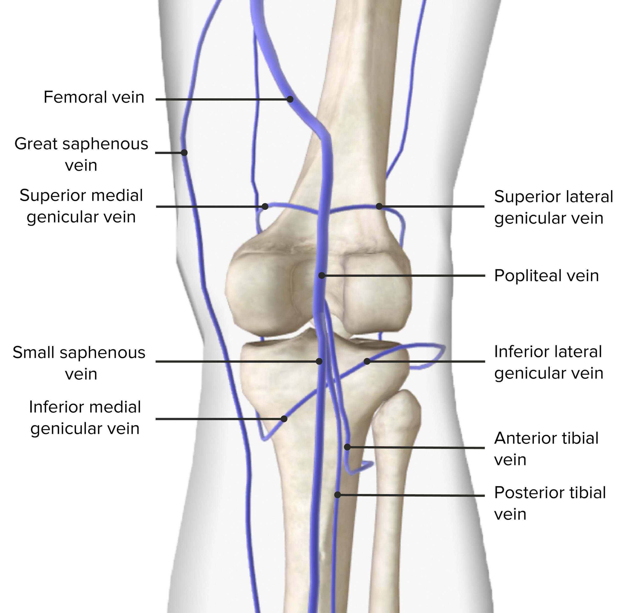
From an anatomical point of view, when the popliteal vein ascends through the fossa, this type of vein will cross at the back of the popliteal artery so that this vein will be located on its lateral side. This popliteal vein will later pass through the adductor hiatus to become the primary part of the femoral vein. The popliteal vein at the level of the popliteus muscle is formed from all the deep veins of the lower leg that unite including the peroneal vein, and the anterior tibial and posterior tibial veins. Furthermore, known as a vein that empties into the popliteal vein, the small saphenous vein, a superficial vessel that drains the posterior part of the lower leg.
LOCATION
The popliteal vein is located in an anatomical area known as the popliteal fossa. The popliteal fossa is a diamond-shaped or shallow depression at the back of the knee joint. This area is bounded by two muscles, namely the semitendinosus and semimembranosus proximomedially, proximolaterally by the biceps femoris, and distally by the two heads of the gastrocnemius muscle. If viewed anatomically, the components located within the popliteal fossa area, from the inside (medially) to the outside (laterally) are the popliteal artery, popliteal vein, and the last one is the tibial nerve. At the time after leaving the popliteal fossa area, this popliteal vein will pass through the adductor Magnus muscle precisely at the adductor hiatus which will later be renamed the femoral vein.
Here are some of the tributaries in the lower leg that drain the popliteal vein:
- Small saphenous vein: This vein drains from the lateral surface of the leg and then continues to the posterior surface of the leg to finally drain into the popliteal vein.
- Anterior tibial vein: This vein adjoins the anterior tibial artery to drain over the anterior interosseous portion of the foot.
- Posterior tibial vein: This vein has the main task of collecting blood from the ankle joints, soles of the feet, and the muscles of the posterior compartment of the foot.
- Genicular vein: This vein is responsible for draining blood starting from the area located around the knee and ending in the popliteal vein.
- Peroneal vein: It drains with the peroneal artery in the lateral compartment of the leg and receives tributaries from the superficial and soleus veins and ends in the posterior tibial vein.
- Sural vein: This vein is located near the posterior leg and drains into the posterior fibula and tibial veins.
ANATOMICAL VARIANTS
A study that was conducted in 2012 of more than 60 corpses, found that there were about 30% anatomical variants of the popliteal vein. These results are also supported by several subsequent studies:
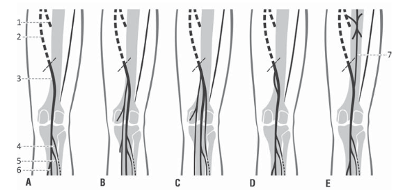
The study conducted by Sadowska et al reported that there were variations in the distribution pattern of the popliteal venous system in the popliteal fossa as follows:
- Single popliteal vein formed in the popliteal fossa;
- High origin of popliteal vein derived from two tributaries;
- High origin of popliteal vein derived from three tributaries;
- The native duplication of a popliteal vein;
- The persistent sciatic vein accompanies the popliteal vein.
Other studies regarding the anatomical variations of the popliteal vein are as follows: 1) The single popliteal vein which is a rare variation reported in about 5% of cases in a study and almost 1% cases of the single persistent sciatic vein found in another study, that is not the popliteal or femoral veins. 2) The course of the vein: there is also a variation of the popliteal vein in direction and/or position perspective compared to the popliteal artery. In some cases, the popliteal vein traverses it either laterally or medially, instead of the typical lateral position and direction of the popliteal artery.
FUNCTION
In general, the popliteal vein has the main function of draining previously deoxygenated blood from the lower legs and then bringing it back to the heart area for oxygenation. Blood flow that is returned to the heart area will be conveyed through a series of venous-muscle pumps. The gastrocnemius pump is also a very important component in this process because when we walk there will be contractions that will encourage direct blood flow to the popliteal vein. In particular, this vein will provide a venous return to the triceps leg or what is known as the gastrocnemius muscle. This muscle is a large, double-headed muscle located in the calf whose main function is to swing the leg back when walking.
ABNORMALITIES
Some anatomical and physiological abnormalities case reports of the popliteal vein are as follows:
- Popliteal Vein Thrombosis: This disorder is in the form of a blood clot or thrombosis that can block the popliteal vein so that there is no blood flow from the lower legs to the heart. Signs and symptoms of thrombosis in the popliteal vein include pain, tenderness, and swelling around the area of the blood clot in the leg.
- Popliteal Vein Entrapment Syndrome: This type of abnormality has been reported in rare cases where the popliteal vein is compressed due to an aneurysm, muscle anomaly, or calf muscle enlargement. This vein will become trapped, making it harder for blood to flow to the leg.
- Popliteal vein aneurysm: The presence of abnormalities that have symptoms of vascular reflux such as swelling, pain, and edema due to trauma that can develop into idiopathic lesions.

 (352 votes, average: 1.09 out of 5)