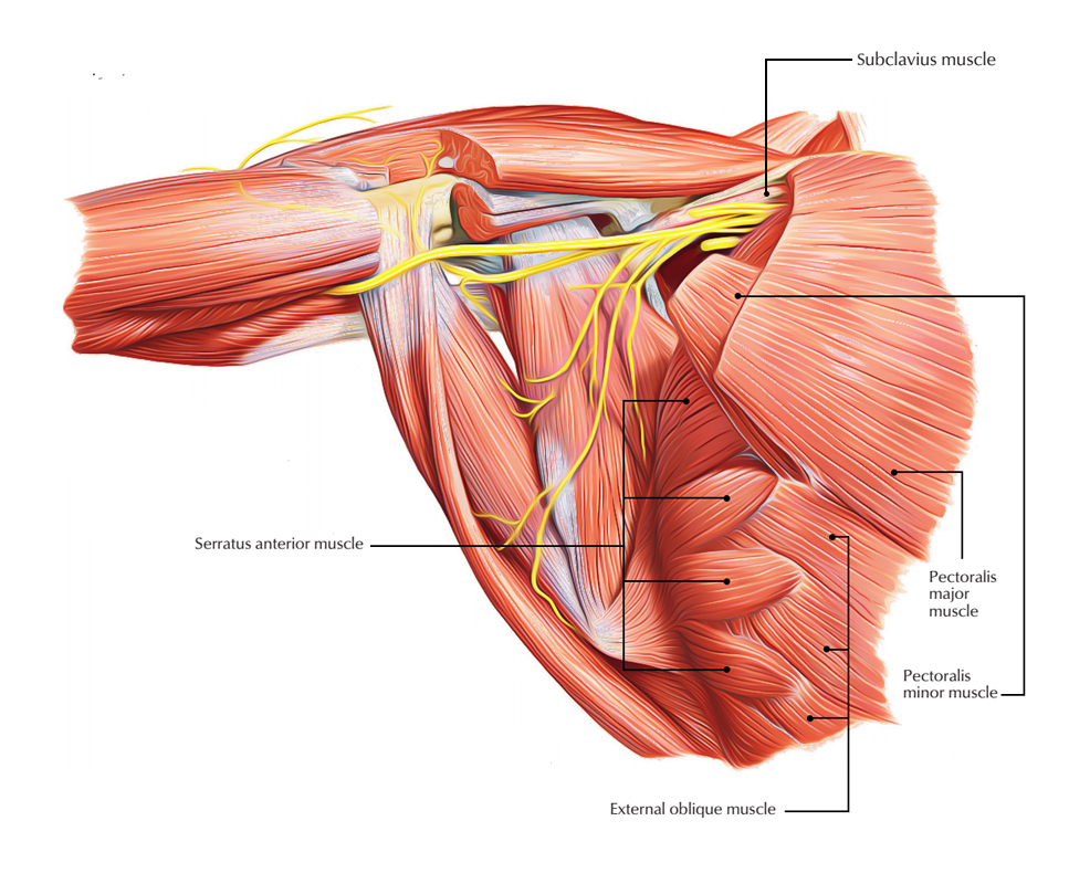
The muscles of the Pectoral Region are:
- Pectoralis major
- Pectoralis minor
- Subclavius
- Serratus anterior
Muscles of the Pectoral Region
The Pectoral region is the one located on the anterior chest wall.
Pectoralis Major
Pectoralis major is the largest muscle of the pectoral region.
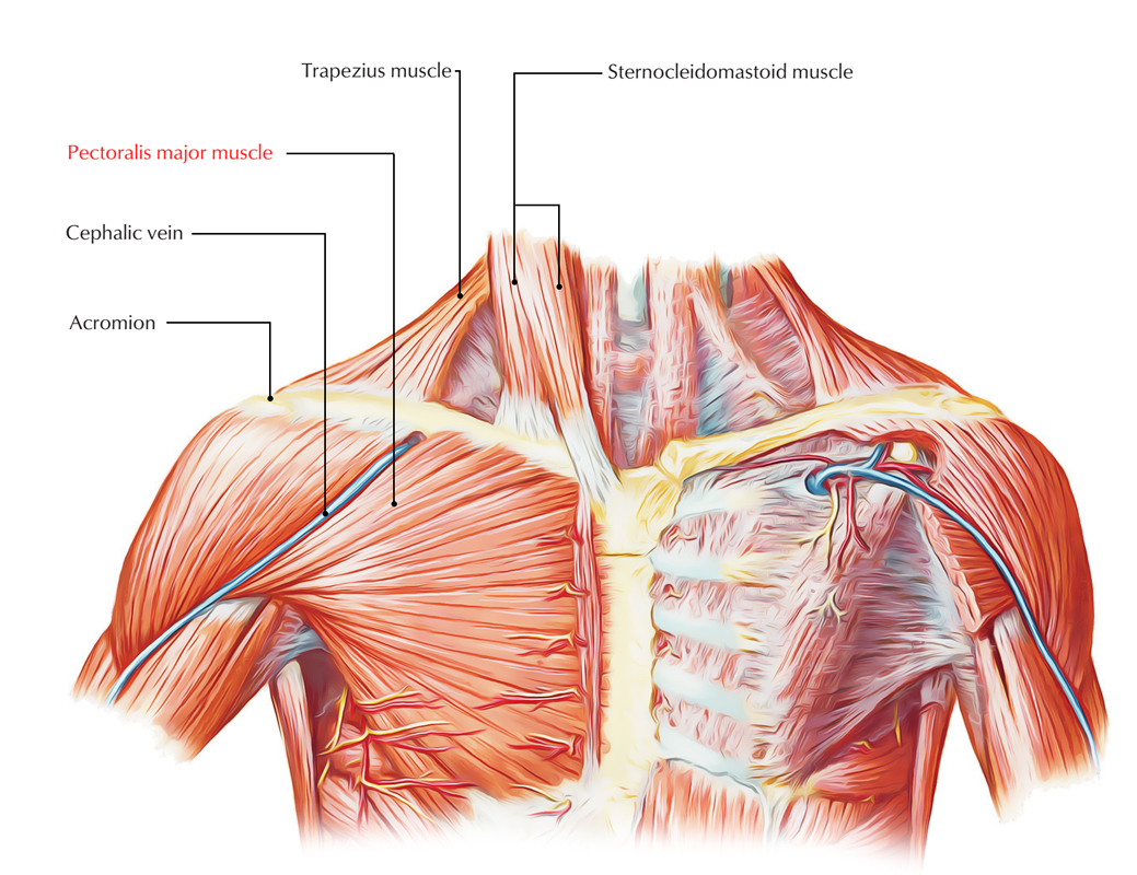
Pectoralis Major Muscle
Origin
Pectoralis major muscle is lean fan shaped and emerges by two heads, viz.
- Small clavicular head.
- Large sternocostal head.
Clavicular head– it arises from the median half of the anterior element of the clavicle.
Sternocostal head– it occurs from the (a) lateral half of the anterior surface of the sternum, up to 6th costal cartilage, (b) median parts of 2nd-6th costal cartilages, and (c) aponeurosis of the external tilted muscle of the abdomen.
Insertion
Pectoralis major is inserted by a U-shaped (bilaminar) tendon on to the sidewise lip of the bicipital groove. The former lamina of the tendon is formed by the clavicular fibres, while posterior lamina is developed by sternocostal fibres. The two laminae are constant with each other inferiorly.
The lower sternocostal and abdominal fibres in their program to insertion are turned in such a way that fibres, which are lowest are put in highest.
This twisting of fibres forms the rounded axillary fold.
Nerve Supply
Nerve supply is by lateral (C5 to C7) and mean pectoral (C8 and Tl) nerves.
- The pectoralis major and pectoralis minor muscles are the only muscles of the upper limb, which are provided by all five spinal segments that form the brachial plexus.
- Sometimes a vertical sheet of muscle fibres prolonging from root of the neck to the top part of the abdomen passes shallow to the medial part of pectoralis major. It is called rectus sternalis/sternalis muscle.
Actions
The clavicular head tightens the arm, while sternocostal head adducts and medially rotates the arm.
- Clinical testing: On lifting a massive rod, the clavicular head becomes prominent and when one tries to lower the rod, the sternocostal head becomes prominent.
Clinical Significance
- Congenital anomaly of pectoralis major: Sometimes, a part of the pectoralis major, normally the sternocostal part, is absent at birth. This results in weakness in adduction and medial rotation of the arm.
Pectoralis Minor
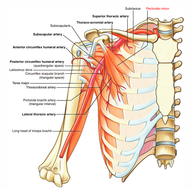
Pectoralis Minor Muscle
It is the small triangular muscle that is located deep to the pectoralis major muscle.
Origin
It ascends from 3rd, 4th, and 5th ribs, near their costal cartilages.
Insertion
It is inserted by a small thick tendon into the median border and upper surface of the coracoid procedure of the scapula.
Nerve Supply
Nerve supply is by medial and lateral pectoral nerves.
Actions
- Assists the serratus anterior in drawing the scapula ahead (protraction) for punching motion.
- Depresses the point of shoulder.
- Acts as an extra muscle of respiration, during forced inspiration.
Important Points
- The pectoralis minor is regarded as the ‘key muscle’ of axilla because it crosses in front of the axillary artery and thus used to split this artery into three parts.
- The origin of pectoralis minor is varied. It may be prefixed (i.e., arises from 2nd to 5th ribs) or postfixed (i.e., arises from 4th to 6th ribs).
- Rarely several fibres of the pectoralis minor split and pass from first rib to the coracoid process to compose what is referred to as pectoralis minimus muscle.
Subclavius
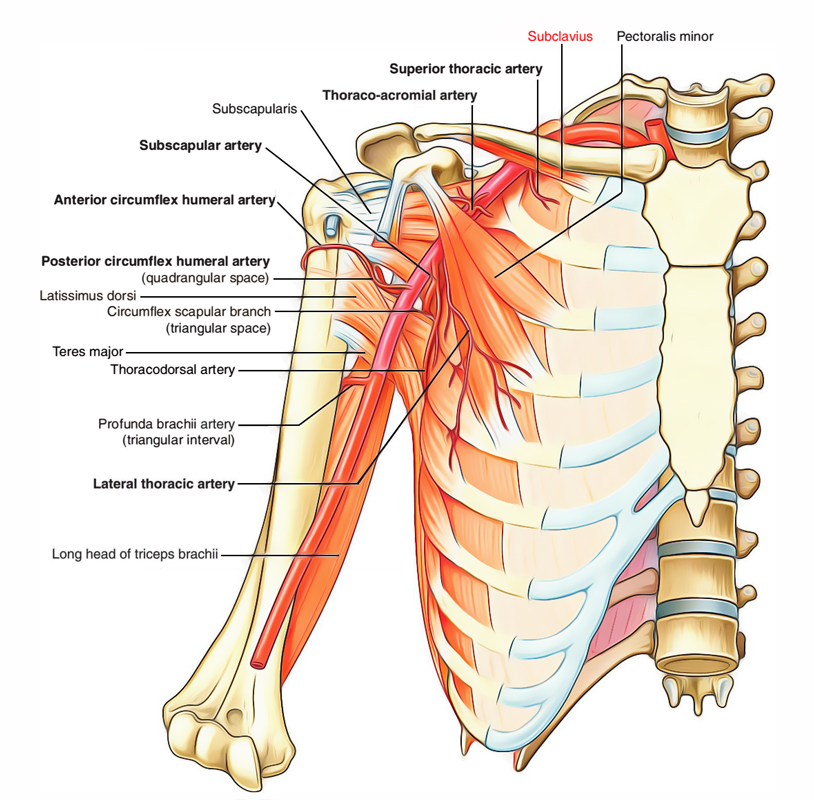
Subclavius Muscle
It is the compact rounded muscle that exists horizontally inferior to the clavicle.
Origin
It arises from the first rib at the costochondral joint.
Insertion
Subclavius is inserted into the subclavian groove on the substandard surface (middle-third) of the clavicle.
Nerve Supply
It is by nerve to subclavius, which emerges from the upper trunk of the brachial plexus.
Actions
The subclavius supports the clavicle by pulling it inferiorly and medially, during action at the shoulder joint.
Serratus Anterior
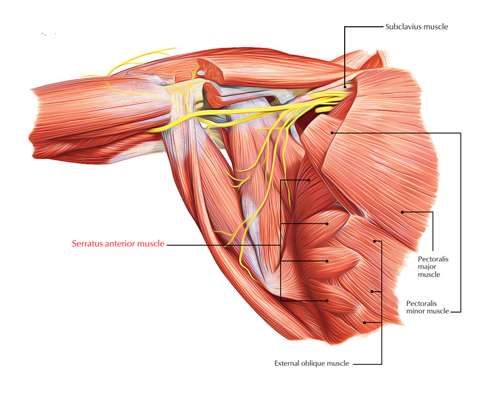
Serratus Anterior Muscle
The serratus anterior is a broad cover of muscle that outfits the side wall of the thorax. Thus purely speaking, it is not a muscle of the pectoral region. But for benefit, it is defined with the muscles of pectoral region.
Origin
It arises by a collection of 8 digitations from upper eight ribs. The first digitation arises from the 1st and 2nd ribs, while all other digitations arise from their equivalent ribs.
Insertion
It is placed into the costal surface of the scapula along its middle border. (The first 2 digitations are inserted into the better angle, next 2 digitations into the medial boundary and the lower 4 or 5 digitations into the inferior angle of the scapula.).
Nerve Supply
It is by lengthy thoracic nerve/nerve to serratus anterior (C5, C6, and C7).
Actions
- It is an effective protractor of the scapula, i.e., it draws the scapula forward about the chest wall for pressing and punching movements as needed during boxing. Hence, serratus anterior is also called boxer’s muscle.
- It keeps the medial/vertebral margin of scapula in firm contact with the chest wall. Its lower 4 or 5 digitations along with lower part of the trapezius revolve the scapula laterally and upwards at the time of overhead abduction of the arm.
Clinical Significance
Paralysis of serratus anterior: The paralysis of serratus anterior muscle adopting an injury to long thoracic nerve by stab injury or during elimination of the breast tumor leads to the following effects:
Protraction of scapula is reduced Inferior angle and middle boundary of scapula become too prominent especially when patient pushes his hands against the wall, producing a clinical situation called winging of the scapula.

 (53 votes, average: 4.53 out of 5)