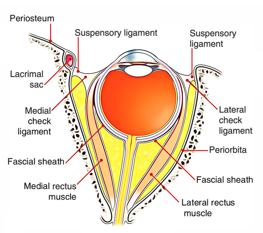
Around every muscle that pierces it, the fascia bulbi gives a tubular sheath. A triangular growth (lateral check ligament) from the fascial sheath of lateral rectus, goes laterally for connection to the lateral wall of the orbit on Whitnall’s tubercle. Likewise, medially for connecting to the medial wall, the orbit on posterior lacrimal crest of the lacrimal bone a triangular growth from sheath of medial rectus (medial check ligament) goes.

Ligaments associated to the Eye
The fascial sheath of inferior rectus thickens on its underside and combinations with the sheath of inferior oblique as well as with the medial and lateral check ligaments creating a hammock-like support for the eyeball. It’s referred to as suspensory ligament of the eye (or suspensory ligament of Lockwood). It’s enlarged in the heart and narrows at its extremities. At the posterior pole of the eyeball the fascia bulbi becomes constant with sheath of the optic nerve.

Ligaments associated to the Eye
Clinical Significance
If suspensory ligament of the eye stays undamaged when the floor of the orbit is fractured or the maxilla is removed surgically, the eyeball doesn’t sag.
During enucleation of the eye, if Tenon’s fascia isn’t damaged and artificial eye is put afterwards inside the socket of Tenon’s capsule it’s capable to move.
- Nerves ofThe Orbit
- Optic Nerve.
- Oculomotor Nerve.
- Trochlear Nerve.
- Abducent Nerve.
- Ophthalmic Nerve.
- Ciliary Ganglion.
Ophthalmic Artery

Eye – Opthalmic Artery
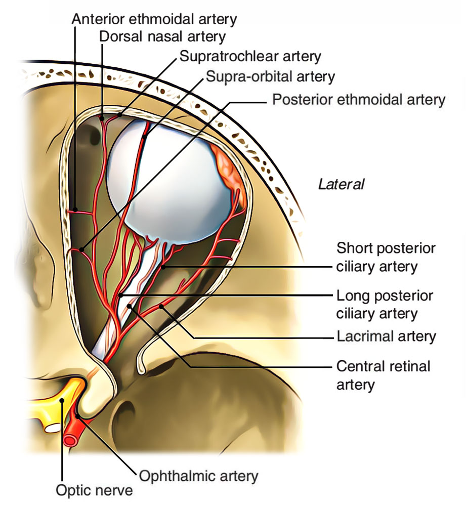
It’s branch of the internal carotid artery. It appears from internal carotid artery as it appears from the roof of the cavernous sinus medial to anterior clinoid process near the optic canal. The artery enters the orbit via optic canal inferolateral to the optic nerve, both being located in a standard dural sheath.
The artery pierces the dura mater, ascends over the lateral side of the optic nerve to cross it superiorly from lateral to medial side together with the nasociliary nerve. It then runs forwards along the medial wall of the orbit tortuously to enable the movements of the eyeball. Near the medial angle of the eye it ends by dividing into 2 branches: supra trochlear and dorsal nasal.
For details see-Opthalmic Artery.
Ophthalmic Veins

Eye – Opthalmic Veins
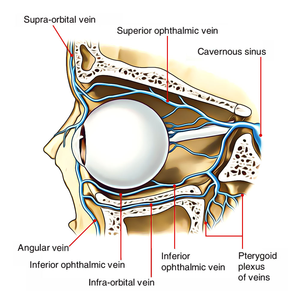
The ophthalmic veins drain the orbit and get tributaries, which correspond to the branches of the ophthalmic artery. The ophthalmic veins are as follows:
Superior ophthalmic vein: It’s a large vein and accompanies the ophthalmic artery. It commences above the medial palpebral ligament and runs backwards above the optic nerve together with the ophthalmic artery, goes through the superior orbital fissure to drain into cavernous sinus.
It interacts anteriorly at its commencement together with the supraorbital and angular veins.
Inferior ophthalmic vein: It runs below the optic nerve and ends either by joining the superior ophthalmic vein or drain directly into the cavernous sinus.
It interacts with pterygoid venous plexus by small veins going through the inferior orbital fissure.
There are not any lymphatics in the eyeball.
Clinical Significance
Because the ophthalmic veins drain into cavernous sinus and convey with all the extracranial veins, they serve as courses via which infection can spread from outside to inside the cranial cavity.
Lacrimal Gland

Eye – Lacrimal Gland
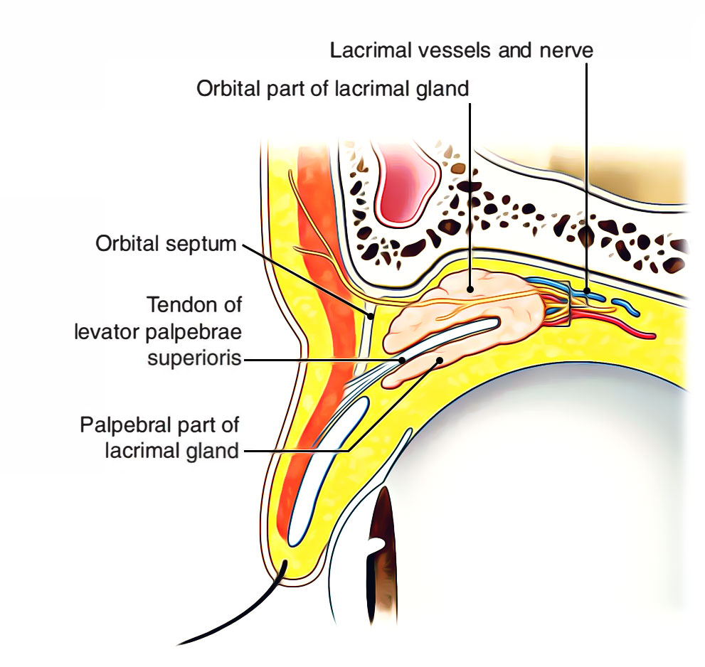
It’s a tubulo-acinar type of exocrine gland, which secretes watery lacrimal fluid. It is composed of 2 parts: orbital and palpebral. The orbital part lies in the lacrimal fossa on the anterolateral part of the roof of the orbit, while palpebral part lies in lateral part of the upper eyelid.
Orbital Fat
It fills up the space between the eyeball, optic nerve and cone of 4 rectus muscles. It acts as a pillow to stabilize the eyeball during its movements.

 (57 votes, average: 4.72 out of 5)