
Orbit
Bounds
The borders of the orbit are as follows:
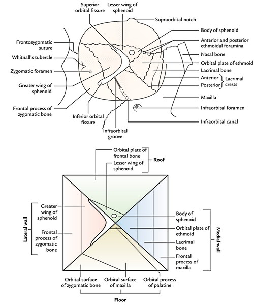
Borders of the Orbit
- Frontal process of maxilla.
- Lacrimal process of maxilla.
- Orbital plate of ethmoid.
- Body of sphenoid.
Lateral wall (most powerful): Created by 2 bones, viz.
- Orbital surface of the zygomatic bone in front.
- Orbital surface of greater wing of sphenoid behind.
Floor: Created by 3 bones:
- Orbital outermost layer of the body of maxilla.
- Orbital surface of the zygomatic bone, anterolaterally.
- Orbital process of the palatine bone, posteromedially.
Roof: Created by 2 bones, viz.
- Orbital plate of the frontal bone in front.
- Lesser wing of the sphenoid behind.
Presenting Features in 4 Boundary Walls of Orbit
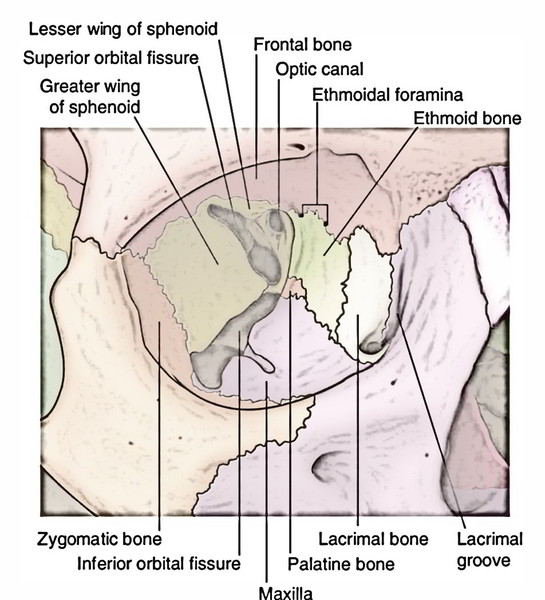
4 Boundaries of the Orbit
- Lacrimal fossa is bounded in front by the anterior lacrimal crest of maxilla and behind by the posterior lacrimal crest of the lacrimal bone. The lacrimal fossa interacts with all the nasal cavity via nasolacrimal canal. The lacrimal fossa and nasolacrimal canal hostel lacrimal sac and nasolacrimal duct, respectively.
- Anterior and posterior ethmoidal foramina, is located in the junction between medial wall and the roof of the orbit.
Lateral wall: Presents 2 features, viz.
- Zygomatic foramen. Occasionally there are 2 small foramina, for zygomaticofacial and zygomatico-temporal nerves.
- Whitnall’s tubercle, a small bony tubercle just behind the lateral orbital margin and somewhat below the frontozygomatic suture.
Floor: Presents 2 features, viz.
- Infraorbital groove and canal.
- Small rough belief in anteromedial angle for origin of inferior oblique muscle.
Roof: Presents 3 features, viz.
- Fossa for lacrimal gland in the anterolateral part.
- Trochlear notch or spine in the anteromedial angle.
- Optic canal at the extreme posterior part of the roofing.
Apex: Is located at the posterior end of the orbit and is composed by sphenoid. More exactly it’s created by the middle of the bony bridge between optic canal and superior orbital fissure. Base: Open and quadrangular, its borders create the orbital margins.
Orbital Margins
The orbital margins are created as follows:
- Supraorbital margin is composed by the frontal bone and presents a notch-supraorbital notch or foramen at the junction of its lateral two-third and medial one-third.
- Infraorbital margin is composed by the zygomatic bone laterally and maxilla medially; it’s constant with anterior lacrimal crest medially.
- Medial orbital margin is ill defined. It’s created by the frontal bone above and lacrimal crest of the frontal process of maxilla below.
- Lateral orbital margin is composed by zygomatic process of the frontal bone above and frontal process of the zygomatic bone below.
The margins of the orbit are easily palpable in vivo. The orbital margins supply a rational bony protection to the eye with the exception of at the lateral margin. For this particular reason protective eye guards are made to compensate for it, in squash and handball players and still allowing great peripheral vision.
Connections
The connections of orbit are as follows:
- Above: Anterior cranial fossa and frontal air sinus (generally).
- Lateral: Temporal fossa in front and middle cranial fossa behind.
- Below: Maxillary air sinus.
- Medial: Ethmoidal air sinuses.
Orbital Fascia or Periorbita
It’s the periosteum of the bony orbit, which lines the bony bounds of the orbit and creates a funnel-shaped fascial sheath that encloses the orbital contents. It’s loosely connected to the bones, for this reason can be easily stripped off especially from roof and medial wall of the orbit.
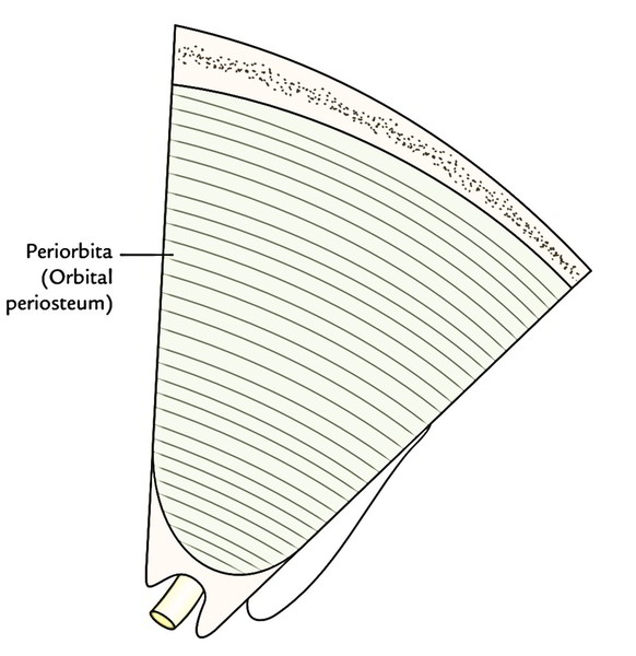
Orbit – Periorbita
Paths to the orbit
There are 3 primary paths by which structures can enter and leave the orbit: – The optic canal transmits the optic nerve and ophthalmic artery. – The superior orbital fissure transmits the lacrimal, frontal, trochlear (CN IV), oculomotor (CN III), nasociliary and abducens (CN VI) nerves. It also carries the superior ophthalmic vein. – The inferior orbital fissure transmits the maxillary nerve (a branch of CN V), the inferior ophthalmic vein, and sympathetic nerves. There are other minor openings into the orbital cavity. The nasolacrimal canal, which drains tears from the eye to the nasal cavity, lies on the medial wall of the orbit. Other little openings consist of the supraorbital foramen and infraorbital canal– they bring little neurovascular structures.
Contents
Contents of the orbit contains these structures:
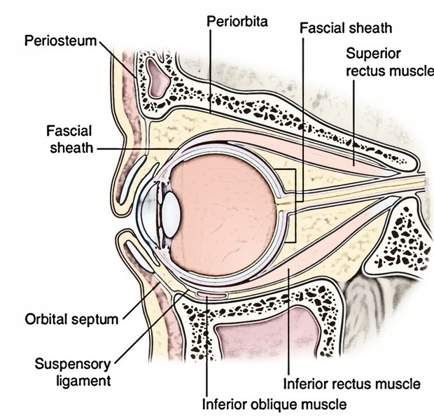
Contents of the Orbit
- Eyeball (most essential content).
- ExtraocularMuscles.
- Fascia bulbi.
- Nerves.
- Optic.
- Oculomotor.
- Trochlear.
- Abducent.
- Ophthalmic.
- Ciliary ganglion.
- Ophthalmic artery.
- Ophthalmic veins.
- Lacrimal gland
- Orbital fat.
All these structures be located inside the orbital periosteum that lines the bony walls of the orbit. Infraorbital nerve and vessels and zygomatic nerve be located outside the orbital periosteum, therefore not contained in the contents of the orbit.
Eyeball (Bulbus Oculi)
The eyeball (L. oculus; Gk. ophthalmos) or ball of the eye is an organ of sight and strongly resembles a camera in its structure. It’s light sensitive retina and is supplied with a lens system (cornea, lens and refractive media) for focusing images and apparatus for controlling the amount of light acknowledged (the iris diaphragm). Additional, such as, for instance, a camera, its interior is black to stop reflection of light. The eyeball is a very lasting structure for its wall enclosing the refractory media, is composed of 3 jackets and the fluid filled up inside it spreads hydraulic pressure uniformly to keep its shape.
Extraocular Muscles
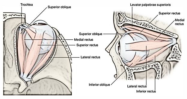
Orbit – Extraocular Muscles
Voluntary Muscles
There are 7 voluntary muscles in the orbit. Of these, 6 muscles move the eyeball and 1 muscle moves the upper eyelid. The muscles moving the eyeball are 4 recti and 2 oblique muscles. The 1 which moves the upper eyelid is referred to as levator palpebrae superioris: 4 recti muscles.
- Superior rectus,
- Inferior rectus,
- Medial rectus and
- Lateral rectus.
2 oblique muscles.
- Superior oblique,
- Inferior oblique.
1 levator palpebrae superioris.
Involuntary Muscles
There are 3 automatic/ smooth muscles, viz. superior tarsal or Muller’s muscle, inferior tarsal and orbitalis.
Fascia BulbiOrFascial Sheath of the Eyeball
The fascia bulbi (Tenon’s capsule) is a free membranous sheath that envelops the eyeball and goes from optic nerve to the sclerocorneal junction. It’s divided from the sclera by the episcleral space. The Tenon’s capsule creates a socket for the eyeball to ease free ocular movements. It divides the eyeball from orbital fat. The fascia bulbi is pierced by:
- tendons of 4 recti and 2 oblique muscles of the eyeball and
- ciliary nerves and vessels around the entry of the optic nerve
Innervation of the Orbit
The innervation of the orbit can be divided into 4 functional elements: basic somatic efferent (extraocular muscles), basic somatic afferent (sensory), basic visceral efferent (autonomic), and special sensory afferent (vision). The basic somatic efferents consist of the motor division of the oculomotor nerve (CN III), the trochlear nerve (CN IV), and the abducens nerve (CN VI).
Venous system
Venous drainage of the orbit takes place through 2 major veins, the superior and inferior ophthalmic veins.
Arterial Supply of the Orbit
Infraorbital artery and Ophthalmic artery with its branches provide the orbit.

 (50 votes, average: 4.60 out of 5)