There are 3 joints between the tibia and fibula: (a) superior tibiofibular, (b) middle tibiofibular, and (c) inferior tibiofibular.
Superior Tibiofibular Joint
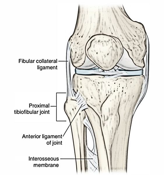
The superior tibiofibular joint is a small plane type of synovial joint between the head of fibula and the lateral condyle of tibia. It might convey together with the knee joint through the popliteal bursa. It allows some amount of gliding or rotatory movements for correcting the lateral malleolus during movements at the ankle joint (example, during dorsiflexion).
Middle Tibiofibular Joint
The middle tibiofibular joint is a fibrous joint created by the interosseous membrane attaching the interosseous edges of the shafts of tibia and fibula. Its fibres are pointed downward and laterally. It’s wide on the upper side and narrow on lower side where it combines with all the interosseous ligament of the inferior tibiofibular joint. A large opening above the upper free margin of interosseous membrane gives passage to the anterior tibial vessels. The interosseous membrane presents a small opening near its lower end for the passage of perforating branch of the peroneal artery.
Functions of The Interosseous Membrane
These are:
- Gives added surface for the connection of muscles.
- Binds tibia and fibula.
- Resists downward movement of fibula by the strong fibular muscles.
Most fibres of the interosseous membrane (vide supra) are pointed downward and laterally with the exception of in the upper part where they’re pointed downward and medially.
Inferior Tibiofibular Joint
The inferior tibiofibular joint is a syndesmosis variety of the fibrous joint. It’s the most powerful of all the 3 tibiofibular joints, since the strength of ankle joint mainly is dependent upon its ethics.
The roughened opposed surfaces of the lower ends of tibia and fibula are joined by an extremely powerful interosseous ligament, which creates the primary bond of union between the lower ends of these bones. The interosseous ligament is covered both in front and behind by the anterior and posterior tibiofibular ligaments, respectively. The posterior tibiofibular ligament is more formidable in relation to the anterior tibiofibular ligament. Its lower and deep portion creates the inferior transverse tibiofibular ligament, that is a solid thick band of yellowish elastic fibres passing transversely from the upper part of malleolar fossa to the posterior border of the articular surface of tibia. It accentuates the concavity of the tibiofibular mortise of the ankle joint.
The inferior tibiofibular joint allows little movements, to enable the lateral malleolus to rotate laterally during dorsiflexion of the ankle.
Comparison of The Superior, Middle, And Inferior Tibiofibular Joints
Superior tibiofibular joint | Middle tibiofibular joint | Inferior tibiofibular joint |
|---|---|---|
| Permits some gliding movements | Permits rotation of fibula during dorsiflexion of the knee | Accentuates the “tibiofibular mortise,” and allows lateral rotation of the lateral malleolus during dorsiflexion |
| Formed by articulation between the oval articular facet on the lateral condyle of tibia and similar facet on the head of fibula | Formed by the union of interosseous border of tibia and fibula by an interosseous membrane | Formed by the union of rough triangular surface of the lower ends of tibia and fibula by a strong interosseous, and anterior and posterior tibiofibular ligaments |
| Innervated by the nerve to popliteus and recurrent genicular nerve | Innervated by the nerve to popliteus | Innervated by the deep peroneal, tibial, and saphenous nerves |
| Plane type of the synovial joint | Fibrous joint | Syndesmosis variety of the fibrous joint |
Test Your Knowledge
Tibiofibular Joint

 (46 votes, average: 4.87 out of 5)