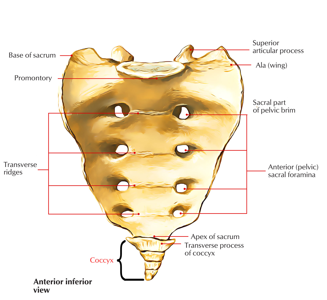
Coccyx or tailbone is a small, triangular bone created by the fusion of 4 vestigial coccygeal vertebrae, which increasingly reduce in size from above downward. At the level of the upper border of the pubic symphysis, the tip of coccyx is located. In the natal cleft, the coccyx can be palpated.

Coccyx
- The word “coccyx” is originated from the Greek word “cuckoo,” a fowl. The tailbone was thus referred to as because its shape resembled the fowl’s beak.
- Morphologically coccyx composes the tail (tailbone).
- The number of coccygeal vertebrae might differ from 3 to 5.
General Features
The coccyx is pointed downward and forward, making a constant curve with the sacrum. It presents these features:
- Base.
- Apex or tip.
- Pelvic surface.
- Dorsal surface.
- Lateral margins.
Special Features
Base
- It’s created by the upper surface of the body of the very first coccygeal vertebra.
- It articulates with the apex of sacrum to create a cartilaginous sacrococcygeal joint.
- A pair of bony processes named coccygeal cornu project from the posterolateral aspects of the body of the very first coccygeal vertebra and are joined with the sacral cornua by the intercornual ligaments.
- The fifth sacral nerves after coming from the sacral hiatus pass laterally via the period between the body of the fifth sacral vertebra and the intercornual ligaments.
- The vestigial transverse processes of the very first coccygeal vertebrae project laterally and upward from the side of the base.
Apex
- It’s created by the body of the fourth coccygeal vertebra.
- It is located about 4 cm behind and above the anal orifice.
- It gives connection to sphincter ani extern us and anococcygeal ligaments.
Pelvic Surface
- It’s related to the rectum.
- It gives connection on every side to the coccygeus muscle in the upper 2 sections and levator ani in the lower 2 bits of the coccygeal vertebra.
- The ganglion impar, where the sympathetic trunks of 2 sides meet, is located on the pelvic surface of first coccygeal vertebra deep to the rectum.
- Anastomosis between the median sacral and inferior lateral sacral arteries and coccygeal body is located on the pelvic surface distal to ganglion impar.
Dorsal Surface
- It gives origin on every side to gluteus maximus.
- It gives origin to sphincter ani externus at the tip.
- It gives connection to filum terminale on the very first coccygeal vertebra.
Lateral Margins
- They give connection to sacrotuberous and sacrospinousligaments.
Ossification
Initially the coccyx begins as a mass which is attached at the end of the sacrum then slowly ossifies as a individual bone. The coccyx ossifies from 4 primary centers, 1 for each segment.
Clinical Significance
Coccydynia
It’s a clinical condition characterized by pain in the coccygeal region. It happens because of separation of coccyx from sacrum following a drop directly on the coccyx.
Spermatic Cord
It’s a soft rounded cord within the males. It can be palpated via skin as it enters downward just above the medial end of inguinal ligament to go into the scrotum.
Mcburney’s Point
It’s a point in the junction of medial 2/3rd and Lateral 1/3rd of the line going from the umbilicus to the anterior superior iliac spine (spinoumbilical line). The base of appendix is located deep to this point.
Murphy’s Point
It’s a point where linea semilunaris meets the right subcostal margin. It corresponds to the tip of the 9th costal cartilage. The fundus of gall bladder is located deep to this point.
Abdominal Planes
Transpyloric Plane/Addison’s Plane
It’s an imaginary horizontal plane which goes through the midpoint of the line joining the jugular (suprasternal) notch to the symphysis pubis. It is located about 1 hand’s width below the xiphisternal joint. Anteriorly it goes through the tip of the 9th costal cartilage and posteriorly via the lower border of the body of L1 vertebra. It’s the key plane of the abdomen as it corresponds to numberof abdominal viscera, viz. pylorus of stomach, hila of kidneys, etc.
Subcostal Plane
It’s an imaginary horizontal plane, which enters immediately below the costal margins. It enters anteriorly via the bottom edges of costal cartilages of the 10th rib, and posteriorly via the body of L3 vertebra.
Transumbilical Plane
It’s a transverse plane that goes through the umbilicus (or navel) and is located at the level of intervertebral disc between the L3 and L4 vertebrae.
Intertubercular Plane
It’s an imaginary horizontal plane which joins the tubercles of the iliac crests. It’s palpable 5 cm posterior to the anterior superior iliac spines and goes through the upper part of the body of L5 vertebra.
Left And Right Vertical Planes (Mid clavicular Planes)
They pass from the midpoint of the clavicle superiorly to the point midway between the anterior superior iliac spine and the pubic symphysis inferiorly, i.e., midinguinal point.
These planes are utilized to delineate the 9 regions or 4 quadrants of the abdominal cavity, which the clinicians send, to explain the location of the abdominal viscera and pain abdomen.
Test Your Knowledge
Coccyx

 (48 votes, average: 4.52 out of 5)