The Abdominal Cavity is the largest body cavity which is present in the torso of the mammals between the thoracic cavity. A central gut tube (gastrointestinal system) which is suspended from the posterior abdominal wall and also somewhat from the anterior abdominal wall with thin sheets of tissue is the typical organization of the abdominal cavity.
- A ventral (anterior) mesentery for proximal regions of the gut tube
- A dorsal (posterior) mesentery along the entire length of the system
Various parts of these two mesenteries are named as per the organs to which they suspend or to which they are associated. Major viscera, such as the kidneys, which are not suspended in the abdominal cavity by mesenteries are associated with the abdominal wall.
The abdominal cavity is lined by peritoneum, which is composed of an epithelial-like single layer of cells (the mesothelium) together with a supportive layer of connective tissue. Peritoneum is similar to the pleura and serous pericardium in the thorax.
The peritoneum reflects off the abdominal wall to become a component of the mesenteries that suspend the viscera.
- Parietal peritoneum lines the abdominal wall.
- Visceral peritoneum covers suspended organs.
Generally, elements of the gastrointestinal tract and its derivatives completely fill the abdominal cavity, making the peritoneal cavity a potential space, and visceral peritoneum on organs and parietal peritoneum on the adjacent abdominal wall slide freely against one another.
Abdominal viscera are either intraperitoneal or retroperitoneal:
- Intraperitoneal structures, such as elements of the gastrointestinal system, are suspended from the abdominal wall by mesenteries
- Structures that are not suspended in the abdominal cavity by a mesentery and that lie between the parietal peritoneum and abdominal wall are retroperitoneal in position
Retroperitoneal structures include the kidneys and ureters, which develop in the region between the peritoneum and the abdominal wall and remain in this position in the adult.
During development, some organs, such as parts of the small and large intestines, are suspended initially in the abdominal cavity by a mesentery, and later become retroperitoneal secondarily by fusing with the abdominal wall.
Boundaries
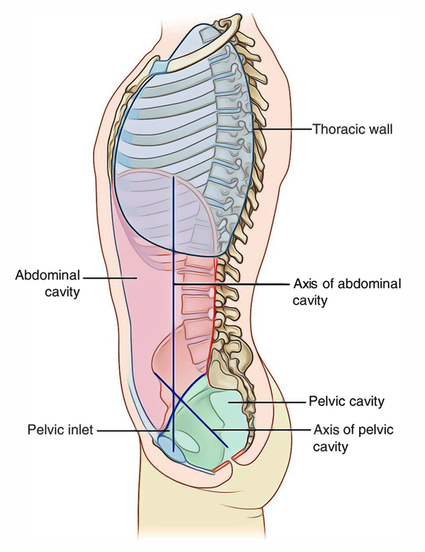
The abdomen is defined as a part of trunk that lies between the diaphragm above and the pelvic inlet below.
The boundaries of the abdominal cavity are as follows:
- Roof: It is formed by the diaphragm, which also forms the upper parts of the lateral and posterior walls.
- Anterior wall: It is made by three pairs of flat muscles (external oblique, internal oblique, and transversus abdominis) and their aponeuroses, and a pair of vertical muscles (rectus abdominis). The vertical muscles lie in the anterior median region, one on each side of the anterior midline, and are enclosed in the aponeuroses of the flat muscles.
- Lateral wall: The upper part of each lateral wall between the ribs and the iliac crest (also called flank) is formed by three flat muscles. The lower part of each lateral wall is formed by the ilium of hip bone covered internally by the iliacus muscle.
- Posterior wall: It is formed by the vertebral column, muscles attached to it (diaphragm, psoas major, and quadratus lumborum), and thoraco-lumbar fascia. Below this, it is formed by the posterior part of ilium and iliacus muscle covering it.
- Floor: It is absent inferiorly as the abdominal cavity communicates with the pelvic cavity at the pelvic brim.
The abdomen and pelvic cavities are lined by a thin serous membrane called peritoneum.
Points to be noticed
- The diaphragm creating the superior boundary moves up and down with respiration.
- The anterior abdominal wall is solid and elastic.
- Firmness of anterior abdominal wall shields the abdominal viscera and its elasticity enables the growth of the abdominal viscera.
- The posterior abdominal wall is osteomuscular and inflexible. Its rigidity gives support to the abdominal organs.
Shape
In transverse section, the abdominal cavity is kidney shaped because the vertebral column protrudes into it posteriorly in the midline. Thus, there is a deep paravertebral gutter on each side of the vertebral column. In median section the abdominal cavity is oblong longitudinally, and its posteroinferior part is continuous with the pelvic cavity.
Contents
The organs and glands of the digestive and urinary systems inhabit majority of the abdominal cavity. These organs and glands are listed below:
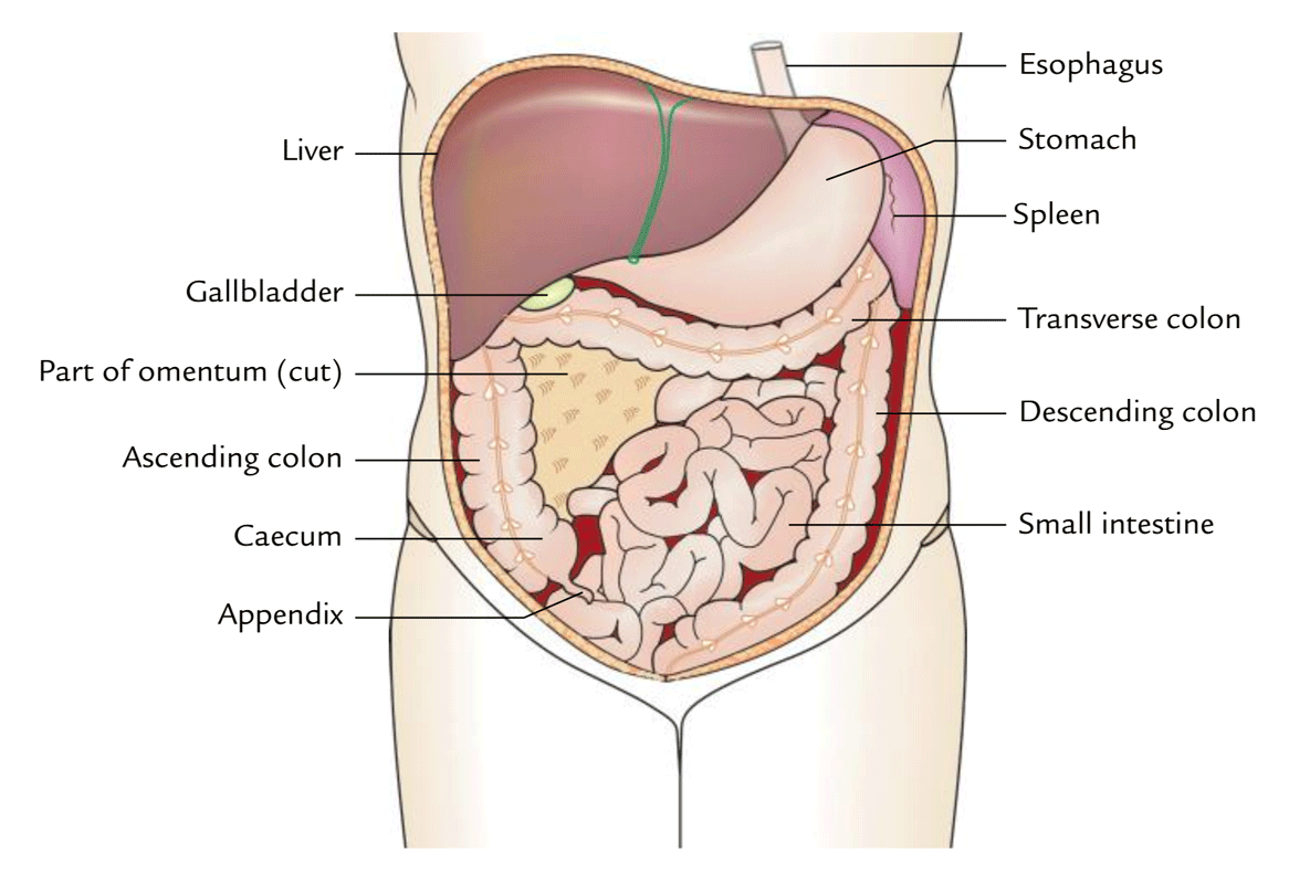
- Stomach, small intestine, and the majority of the large intestine.
- Liver, gallbladder, and pancreas.
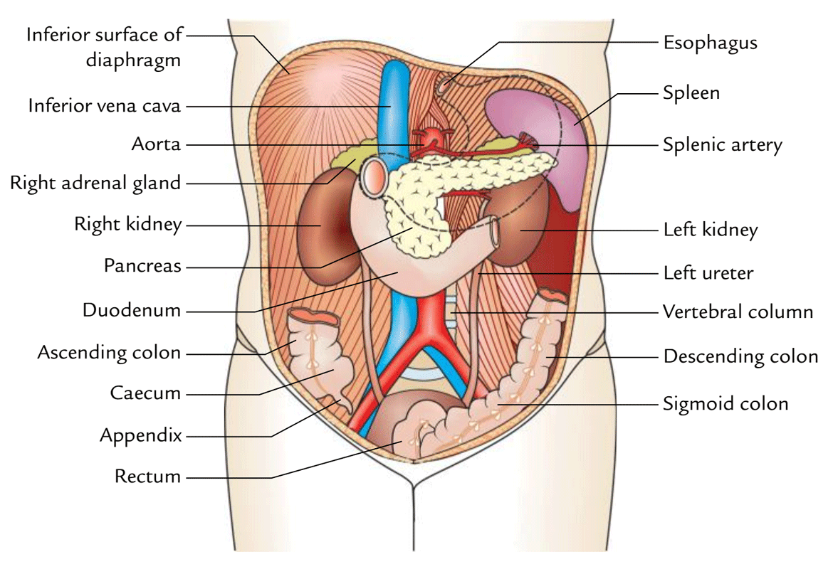
- 2 kidneys and upper part of the ureters.
- Adrenal glands (also referred to as suprarenal glands).
- Other structures contain blood vessels, lymph vessels, nerves, spleen, and lymph nodes.
Organs occupying anterior part of the abdominal cavity.
Organs occupying posterior parts of the abdominal cavity
Nine Regions of the Abdominal Cavity
The clinicians divide the abdominal cavity into nine regions to describe the location of abdominal organs and the pain associated with them during physical examination.
The abdominal cavity is divided into nine regions by four imaginary planes (two vertical and two horizontal) on the anterior abdominal wall.
1. Superior horizontal plane: It corresponds to the transpyloric plane of Addison. It is placed midway between the suprasternal notch and the pubic symphysis. This however is an awkward estimation during physical examination for a clinician, and a simpler and practical method is to locate a point midway between the umbilicus and the lower end of the body of sternum. The xiphoid process should not be used because its size is variable. The transpyloric plane lies at the level of lower border of LI vertebra and cuts the costal margin at the 9th costal cartilages.
Occasionally the subcostal plane is used in preference to the transpyloric plane. This is drawn through the lowest parts of the costal margins at the 10th costal cartilages and lies at the level of the body of L3 vertebra.
2. Inferior horizontal plane (intertubercular plane): It is drawn at the level of tubercles of the iliac crests, which are palpable 5 cm posterior to the anterior superior iliac spine. The intertubercular plane lies at the level of upper border of L5 vertebra.
3. Right and left vertical planes (midclavicular planes): Each vertical plane passes vertically downward from the midpoint of the clavicle to the midinguinal point (a point midway between the anterior superior iliac spine and the pubic symphysis).
Nine regions thus marked out are arranged into three horizontal zones of abdomen: upper, middle, and lower. From right to left, in the upper abdomen they are designated as right hypochondrium, epigastric region, and left hypochondrium. In the middle abdomen they are designated as right lumbar region, umbilical region, and left lumbar region. In the lower abdomen they are designated as right iliac fossa, hypogastric (pubic) region, and left iliac fossa.
Regions of the Abdomen
| Zone | Regions (right to left) |
| Upper abdomen | Right hypochondrium, epigastric region, left hypochondrium |
| Middle abdomen | Right lumbar region, umbilical region, left lumbar region |
| Lower abdomen | Right iliac fossa, hypogastric region (pubic), left iliac fossa |
Abdominal Regions and their Main Contents
| Region | Contents |
| Right hypochondrium | • Liver • Gallbladder |
| Epigastric region | • Stomach • Pancreas • Duodenum |
| Left hypochondrium | • Spleen • Left colic flexure |
| Right lumbar region | • Right kidney • Right ureter • Ascending colon |
| Umbilical region | • Loops of small intestine • Aorta • Inferior venacava |
| Left lumbar region | • Left kidney • Left ureter • Descending colon |
| Right iliac fossa | • Caecum • Appendix |
| Hypogastric region | • Coils of small intestine • Urinary bladder (if distended) • Uterus (if enlarged) |
| Left iliac fossa | • Sigmoid colon |
Four Quadrants of the Abdominal Cavity
For more general clinical descriptions, the abdominal cavity is divided into four quadrants by a horizontal transumbilical plane passing through the umbilicus and a vertical median plane intersecting the horizontal plane at the umbilicus. The four quadrants thus formed are:
- Right upper quadrant.
- Left upper quadrant.
- Right lower quadrant.
- Left lower quadrant.
Abdominal Viscera
The abdominal viscera contains stomach and intestines, their affiliated glands (liver and pancreas), blood and lymph vessels, spleen, kidney, and suprarenal glands.
Stomach
The stomach is the most dilated part of the digestive tube. It is J-shaped and located in the abdominal cavity below the diaphragm somewhat to the left of the mid-line. The capacity of the stomach is about 1500 ml in the grownup.
Functions
The primary functions of the stomach are as follows:
- Churning and breaking of food and blending it with the gastric juice secreted by specialized glands in its mucosa.
- Keeping the food briefly.
- Secreting intrinsic factor necessary for absorption of vitamin B12.
Small Intestine
The small intestine is a convoluted tube linking stomach together with the large intestine. The length of small intestine is about 6 meter and stretches from the pyloric sphincter to the ileocecal junction. The small intestine lies in the central and lower parts of the abdominal cavity surrounded by the large intestine. It is composed of 3 parts: (a) duodenum, (b) jejunum, and (c) ileum.
The duodenum is proximal short arch (c-shaped) portion and is about 25 cm long. It’s the broadest and most fixed part of the small intestine. The ducts from gallbladder and liver and pancreas goes into it.
The jejunum is the name given to the upper two fifth of the balance of the small intestine and the lower 3-fifth is named ileum.
Functions
The primary functions of the small intestine are digestion of food and absorption of nutriments.
Large Intestine
The large intestine starts at the end of the ileum as caecum and ends at the anus. It’s about 1.5 m long and creates an arch around the coiled up small intestine. For illustrative purposes, it’s split into following 7 parts:
- Caecum and appendix.
- Ascending colon.
- Transverse colon.
- Descending colon.
- Sigmoid colon.
- Rectum.
- Anal canal.
Functions
The primary functions of the large intestine are as follows:
- Absorption of water and salts.
- Formation and excretion of feces.
Liver
The liver is the largest gland of the body. It’s situated in the upper right part of the abdominal cavity. It’s 2 main lobes: left and right. The right lobe is substantially bigger compared to the left lobe.
Functions
A few significant functions of the liver are as follows:
- Metabolism of carb, fat, and protein.
- Detoxification of drugs and toxins.
- Storage of glycogen and fat-soluble vitamins (ADEK).
- Secretion of bile.
Gallbladder
The gallbladder is a pear shaped organ situated on the under surface of the right lobe of the liver. It gets bile from the liver, which it stores and concentrates. When greasy food enters the duodenum, the bile is poured into the intestine via the bile duct by the contraction of the walls of the bladder.
Clinical Significance
Jaundice
It’s a clinical condition characterized by yellowing of the skin and sclera of the eyes. It takes place when bile enters the blood in liver diseases like hepatitis or cirrhosis of the liver where the liver cells break down and release bile into the blood. Bile also enters into the blood when outlet of bile from the gallbladder to intestine via the bile duct is obstructed.
Pancreas
The pancreas is an elongated, soft, light, and delicately lobulated gray gland. Pancreas is located transversely across the posterior abdominal wall and is about 12-15 cm in length. It is composed of broad head, neck, body, and a narrow tail. The head of the gland is located inside the curve of the duodenum, the body supporting the stomach and tail in front of the left kidney. The tail goes as far as the spleen.
Functions
The pancreas is an exo-endocrine gland. The function of exocrine pancreas will be to create pancreatic juice consisting of enzymes that digest carbohydrates, proteins, and fats.
The function of endocrine pancreas is always to secrete hormones, insulin and glucagon, which control the blood glucose level.
Clinical Significance
Diabetes Mellitus
Lack of insulin ends in diabetes mellitus. The blood sugar level rises above the renal threshold and glucose is lost in the urine.
Spleen
The spleen is a large wedge shaped mass of vascular and lymphoid tissue. It’s purplish red in colour and is located high up at the back of the abdominal cavity on the left side supporting the stomach.
Functions
The chief functions of the spleen are as follows:
- Destruction of red blood cells.
- Generation of fresh lymphocytes for the blood stream.
Kidneys
The kidneys are 2 bean-shaped organs situated on the posterior abdominal wall, 1 on every side of the vertebral column, behind the peritoneum. The right kidney generally is located at a somewhat lower level in relation to the left.
Function
The primary function of the kidney would be to secrete and excrete urine.
The makeup of blood must not change beyond specific limits if the tissues of the body are to stay healthy. This regulation is dependent upon the removal of damaging waste products and maintenance of water and electrolyte balance.
Ureters
The ureters are 2 tubes, which attach the kidneys to the urinary bladder. Every ureter is normally 25 cm long with a diameter of about 3 mm.
Function
The ureters transportation urine from the kidneys to the urinary bladder.
Adrenal Glands (Suprarenal Glands)
There are 2 suprarenal glands – left and right. Every gland tops the upper pole of the corresponding kidney. The right gland is triangular on the other hand the left gland is semilunar in shape. Every gland is composed of 2 parts: cortex and medulla.
Functions
The use of the cortex would be to secrete a number of steroid hormones, which are responsible for: (a) maintenance of electrolyte and water balance, (b) maintenance of blood sugar concentration and of liver and muscle glycogen, and (c) control of inflammatory responses.
The function of medulla will be to secrete adrenaline and noradrenaline in the blood, which serve as neurotransmitters.
Clinical significance
The knowledge of structures present in the nine regions helps the clinician to know the source of pain. The general guidelines are as follows:
- Pain in the right hypochondrium comes from the gallbladder and biliary ducts.
- Pain in the epigastric region comes from the stomach and duodenum.
- Pain in the left hypochondrium comes from the pancreas.
- Pain in the right lumbar region comes from the right kidney.
- Pain in the umbilical region comes from the small intestine.
- Pain in the left lumbar region comes from the left kidney.
- Pain in the right iliac fossa comes from the vermiform appendix.
- Pain in the hypogastrium comes from the urinary bladder and uterus.
- Pain in the left iliac fossa comes from the sigmoid colon. It is important to note that pain in the abdomen usually occurs due to two reasons: (a) inflammation, and (b) obstruction of conducting muscular tubes such as the bowel or the ureter.

 (61 votes, average: 4.33 out of 5)