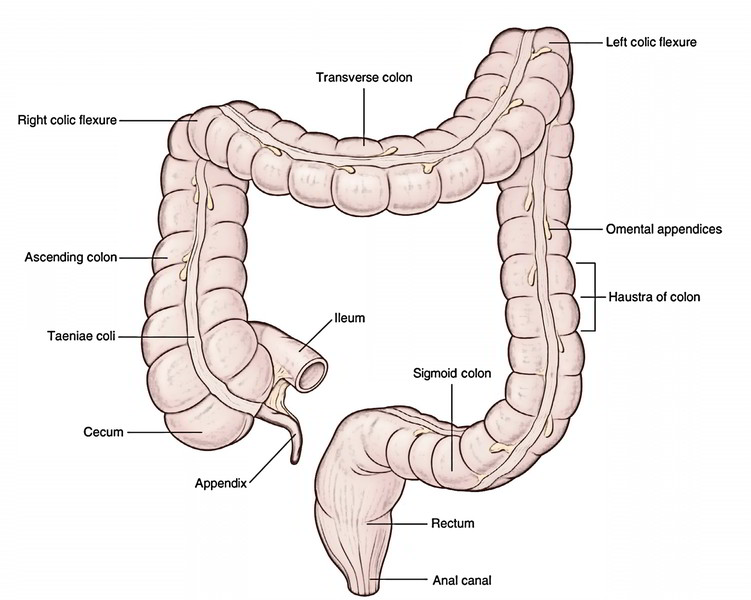
The Large Intestine is amde up of caecum, colon, rectum and anus. The length of the large intestine goes about 1.5 m. It starts from the caecum to its right, iliac fossa to the anus in the perineum.
As compared to the small intestine, it is more fixed in position, aside from the transverse colon and sigmoid colon.
The large intestine performs the following functions:
- Absorption of water from fluid contents in it to help create the feces.
- Storage, lubrication, and expulsion of feces.
- Synthesis of vitamin B complex by normal bacterial flora present its lumen.
- Protection from invasion by microorganisms by its mucoid secretion that is rich in IgA group of antibody.
Parts
For illustrative purposes, the large intestine is split into the following 4 parts:
- Caecum and appendix.
- Colon.
- Rectum.
- Anal canal.
The colon is further split into 4 parts: ascending colon, transverse colon, descending colon, and sigmoid colon.
All the parts of the large intestine are retroperitoneal and mended with the exception of appendix, transverse colon, and sigmoid colon that are intraperitoneal and possess mesenteries. The mesenteries of these parts are called mesoappendix, transverse mesocolon, and sigmoid mesocolon, respectively.
Structure
The large intestine gets its name because its diameter (6.5 cm; 2.5 in) is larger than that of the small intestine, although its length (1.5 m; 5 ft) is much shorter. The large intestine consists of four segments: cecum, colon, rectum, and anal canal.
The first portion of the large intestine is the pouchlike cecum, which bulges inferior to the ileal orifice. The slender, wormlike appendix extends from the cecum and, although it has no digestive function, it contributes to the immune defense of the body.
The colon forms most of the large intestine and is subdivided into four segments. The ascending colon extends superiorly from the cecum along the right side of the abdominopelvic cavity. As it nears the liver, it turns left to become the transverse colon. Near the spleen, the transverse colon turns inferiorly to become the descending colon along the left side of the abdominopelvic cavity. Near the pelvis, the descending colon becomes the sigmoid colon, which is characterized by an S-shaped curvature leading to the rectum.
The rectum is the straight portion of the large intestine that continues inferiorly from the sigmoid colon through the pelvic cavity and ends at the anal canal. The anal canal is the last 3 cm of the large intestine and its external opening is the anus. The mucosa of the anal canal is folded to form the anal columns, which contain networks of arteries and veins. The anus is kept closed except during defecation by the involuntarily controlled internal anal sphincter and the voluntarily controlled external anal sphincter.
The colon has a puckered appearance when viewed externally. This results because the longitudinal muscles are not uniformly layered but are reduced to three longitudinal bands, the taeniae coli, that run the length of the colon. Contraction of the taeniae coli gathers the colon into a series of pouches called haustra. Like the small intestine, the large intestine is supported by a mesentery.
The mucosa of the large intestine is also different from that of the small intestine. Villi are absent, and the simple columnar epithelium contains numerous mucus- producing goblet cells.
Mucosa
The mucosa will not present transverse circular folds (plicae circulares) and villi. Nonetheless, temporary folds affecting mucosa and submucosa exist in the undescended colon. The surface epithelium is absorptive in nature and is composed of columnar cells bearing microvilli.
Intestinal glands (crypts of Lieberkuhn) are long and tubular and possess a large number of goblet cells. The lamina propria of the mucous membrane includes ample diffuse lymphatic tissue but is devoid of Peyer’s patches.
Submucosa
It’s created from loose areolar tissue and includes blood vessels, lymph vessels, and nerve plexus (Meissner’s plexus).
Muscle Layer
It contains outer longitudinal and inner circular layers of smooth muscle. The outer longitudinal layer is not continuous, instead, it’s ordered into 3 thick longitudinal bands referred to as teniae coli. The inner circular layer of smooth muscle Teniae coli is thin when compared with the small intestine.
Serosa
It covers the transverse colon and the sigmoid colon but the parts of ascending and descending colons are covered by tunica adventitia.
Functions
Chyme residue entering the large intestine contains water, minerals, bacteria, and other substances that were not digested or absorbed while in the small intestine. There are no digestive enzymes secreted by the large intestine.
Instead, intestinal bacteria decompose the undigested food molecules. This action yields certain B vitamins and vitamin K, in addition to gas (flatus). The mucosa of the large intestine secretes large quantities of mucus that lubricate the intestinal lining and reduce abrasion as materials are moved along.
A major function of the large intestine is the absorption of water, some minerals, and vitamins as the contents slowly move through the colon. Much of this absorption occurs before the chyme reaches the descending colon, where it is congealed to form the feces (fe-sez). Feces contain large amounts of bacteria, mucus, and water as well as undigested food molecules.
Cardinal (Distinguishing) Features
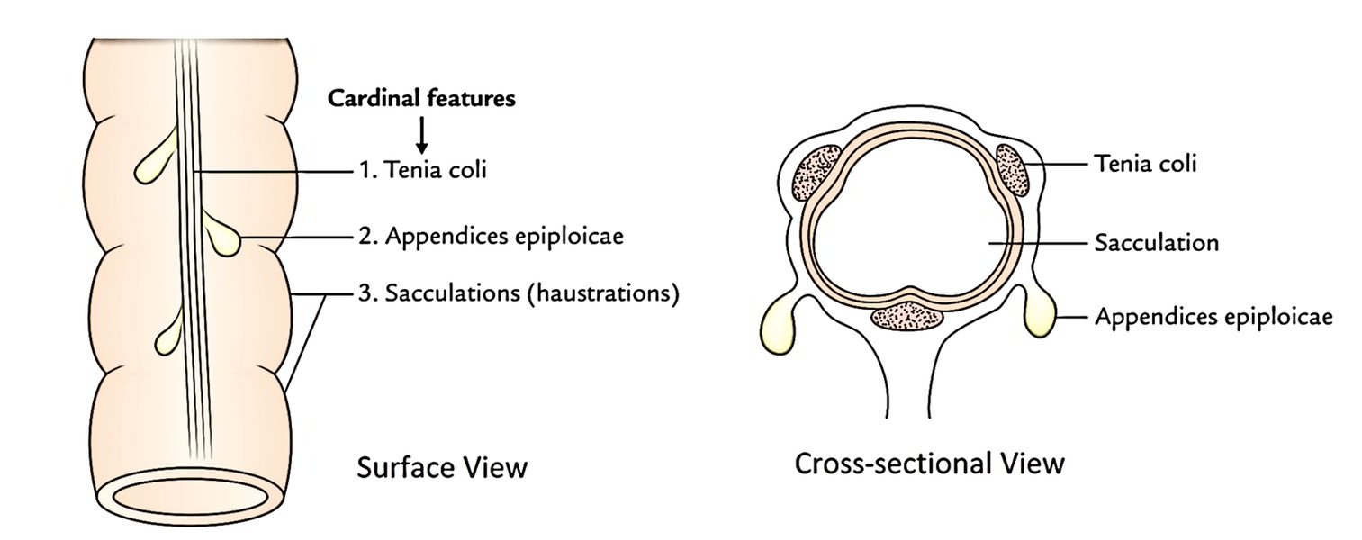
The 3 cardinal features of the large intestine are the presence of (a) teniae coli, (b) appendices epiploicae, and (c) sacculations (or haustrations).
Teniae Coli
These are 3 ribbon-like bands of the longitudinal muscle layer. These bands converge proximally at the base of the appendix and spread out distally to become constant with the longitudinal muscle layer of the rectum. Hence, teniae coli are present on all the parts of colon and caecum.
Location
In the caecum, ascending colon, and descending colon, the places of teniae are anterior (Teniae libera), posteromedial (Teniae mesocolica), and posterolateral (Teniae omentalis), however in the transverse colon the corresponding places are inferior, posterior, and superior, respectively.
Appendices Epiploicae
All these are small bags of visceral peritoneum filled up with fat connected to the teniae of large intestine. Consequently, they may be absent in the appendix, rectum, and anal canal. The appendices epiploicae are most numerous on the sides of sigmoid colon and posterior surface of the transverse colon.
Sacculation (or Haustrations)
All these are a series of pouches/dilatations in the wall of caecum and colon between the teniae. They may be generated because length of teniae fall short of the length of circular muscle coat. The sacculations are accountable for the characteristic puckered look of the large intestine.
Movements
Segmentation and peristalsis within the large intestine are more sluggish than those of the small intestine. Vigorous peristalsis occurs only two to four times a day, usually following a meal. These peristaltic contractions are called mass movements because they move the contents of the descending and sigmoid colons toward the rectum. The defecation reflex is activated when the rectum fills with feces and its wall is stretched. Parasympathetic nerve impulses stimulate muscular contractions that increase pressure within the rectum and relax the internal anal sphincter. Defecation, or expulsion of feces, occurs if the external anal sphincter is voluntarily relaxed. If its contraction is voluntarily maintained, defecation is postponed.
Differences between the Small and Large Intestines
| Features | Small intestine | Large intestine |
|---|---|---|
| Length | 6 m | 1.5 m |
| Lumen | Narrower | Wider |
| Mobility | More | Less |
| Transverse mucous folds | Permanent and not obliterated by distension of the gut | Temporary and obliterated by distension of the gut |
| Villi | Present | Absent |
| Peyer’s patches | Present | Absent |
| Appendices | Appendices | Appendices |
| epiploicae | Absent | Present |
| Teniae coli | Absent | Present |
| Sacculation | Absent | Present |
Medically, it’s essential for the pupils to understand that (a) the small intestine is a standard site for worm infestation, typhoid, and tubercular ulcers on the other hand the large intestine a standard site for amebiasis and carcinoma. (b) The infection and irritation of the small intestine result in diarrhea, on the other hand the infection and irritation of the large intestine cause dysentery.


 (177 votes, average: 2.45 out of 5)